

You can protect your skeletal strength and avoid devastating fractures by recognizing subtle physical indicators long before standard screening milestones arrive. While osteoporosis frequently progresses without obvious discomfort, identifying early signs of bone loss enables you to take immediate control through focused nutrition, progressive resistance loading, and targeted diagnostic scans. Paying close attention to physical shifts—including unexplained reductions in grip strength, receding gum tissue, or gradual height loss—gives you a vital window to collaborate with your physician. Intervening early preserves your bone mineral density, safeguards your mobility, and secures your physical independence throughout every stage of life.
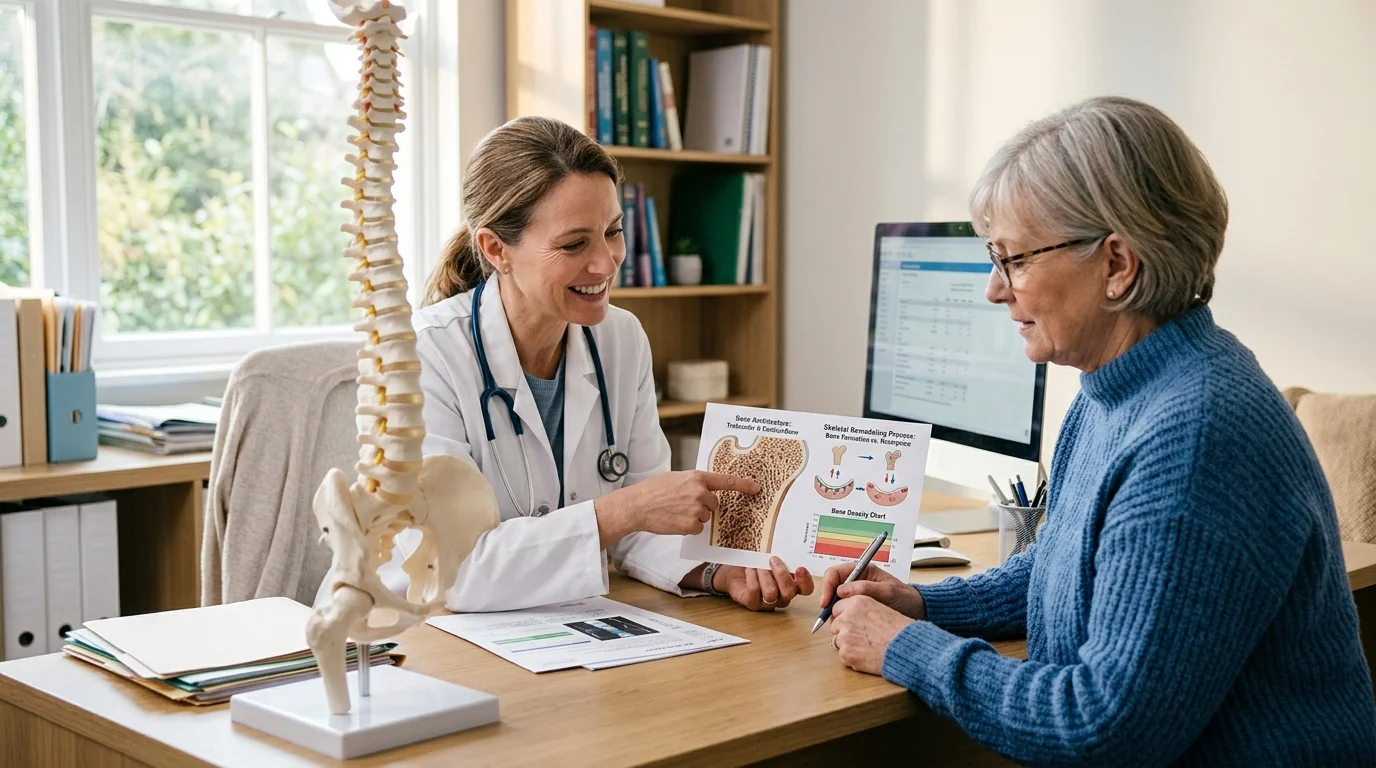
Understanding Bone Density and Silent Skeletal Remodeling
Your bones remain in a constant state of living renewal, undergoing continuous breakdown by specialized cells called osteoclasts and rebuilding by cells known as osteoblasts. During your twenties, osteoblastic activity outpaces breakdown, allowing you to reach peak bone mass around age thirty. After this physiological peak, resorption gradually exceeds formation; if the balance tips too far, the internal architecture of your bones becomes porous, thin, and vulnerable to sudden fracture.
Medical professionals evaluate this internal structure through bone mineral density testing, expressing your score relative to a healthy young adult reference range. According to World Health Organization criteria, a T-score of -1.0 or higher represents normal bone density, while a score between -1.0 and -2.5 indicates osteopenia, or low bone mass. A T-score of -2.5 or below defines clinical osteoporosis, and a score of -2.5 or lower paired with a history of a fragility fracture indicates severe osteoporosis.
According to Centers for Disease Control and Prevention surveillance data, approximately 10.2 percent of adults over age fifty currently live with osteoporosis, a figure projected to climb to roughly 13.6 percent by 2030. Lifetime risk calculations show that approximately fifty percent of women and up to twenty to twenty-five percent of men over fifty will sustain an osteoporotic fracture. Because bone thinning produces no generalized aches in its initial stages, recognizing early structural and systemic markers is vital to preserving your skeletal longevity.

9 Early Signs of Bone Loss and Osteoporosis
1. Noticeable or Measured Height Reduction
Losing measurable height represents one of the most reliable physical markers of underlying bone demineralization. A documented height loss of two centimeters (approximately 0.75 to 0.8 inches) over a short interval, or a cumulative loss of four to six centimeters (1.5 to 2.5 inches) from your peak young-adult height, serves as a primary clinical red flag. This vertical compression occurs when weakened vertebral bodies collapse under the normal downward force of gravity, leading to micro-fractures in the spine. Clinical studies demonstrate that approximately two-thirds of these spinal compression fractures remain completely painless, causing silent height loss that goes unnoticed unless you measure your stature regularly.
2. Periodontal Bone Loss and Receding Gums
Your jawbone serves as the foundational anchor for your teeth, and systemic skeletal thinning frequently manifests in oral tissues first. When systemic bone density drops, the alveolar bone surrounding your tooth roots can recede, causing your gum line to pull backward and exposing sensitive root surfaces. Dentists often detect early systemic osteopenia on routine panoramic X-rays before patients experience symptoms elsewhere in the body. If your dental hygienist notes unexplained bone resorption around your teeth or your dentures suddenly feel loose, treat this discovery as an invitation to discuss a comprehensive bone mineral density evaluation with your physician.
3. Unexplained Loss of Handgrip Strength
Physical therapists and geriatricians frequently evaluate grip strength as an accessible proxy for total skeletal and muscular integrity. Clinical research consistently links low handgrip strength measured by a dynamometer to reduced bone mineral density in both the femoral neck and lumbar spine. Because muscle pull directly stimulates bone remodeling through mechanical tension, declining neuromuscular force often runs parallel to accelerated bone resorption. If you notice increasing difficulty opening sealed jars, turning stubborn doorknobs, or carrying standard grocery bags, your musculoskeletal system may be signaling a broader systemic decline in density.
4. Gradual Postural Curvature and Rounded Shoulders
A subtle forward rounding of the upper back—clinically termed dorsal kyphosis and colloquially known as a dowager’s hump—points toward architectural weakness in the thoracic vertebrae. When weakened anterior portions of your vertebrae compress into wedge shapes, your upper spine bows outward, pulling your shoulders forward and tilting your head downward. You may initially notice that your clothes drape unevenly, that your upper back tires quickly when standing, or that resting flat on the floor feels uncomfortable. Addressing this postural evolution early prevents chronic thoracic spine deformation and preserves vital respiratory capacity.
5. Sudden, Localized Spinal Pain
While gradual bone demineralization itself causes no direct nerve stimulation, a sudden spasm or sharp pain along your mid-to-lower spine can signal a silent vertebral compression fracture. This pain often occurs during mundane daily activities that involve minor spinal flexion—such as bending forward to tie your shoes, lifting a moderate package, or stepping down abruptly from a curb. Patients frequently mistake this localized discomfort for a simple muscular strain or sciatica. If a sharp back ache persists beyond a few days without an obvious traumatic injury, ask your clinician for diagnostic imaging to check your vertebral integrity.
6. Sustaining a Low-Trauma Fragility Fracture
Breaking a bone after minimal trauma represents the most definitive clinical sign that your bone quality has deteriorated. Clinicians define a fragility fracture as any fracture resulting from a fall from standing height or less, or from basic daily movements like coughing or turning in bed. Fracturing a wrist, rib, collarbone, or ankle under low-impact circumstances indicates that your internal bone architecture can no longer absorb standard mechanical loads. Experiencing any low-trauma fracture after age fifty warrants immediate diagnostic follow-up rather than assuming the incident was merely bad luck.
7. Early Menopause and Premature Ovarian Insufficiency
Estrogen plays an indispensable role in maintaining bone density by inhibiting the activity of bone-resorbing osteoclasts. When you transition through menopause, circulating estrogen drops sharply, accelerating bone turnover and leading to average bone density losses of one to three percent per year during the initial postmenopausal decade. Experiencing surgical menopause, premature ovarian insufficiency, or natural menopause before age forty-five dramatically lengthens the total duration of estrogen deficiency. This biological timeline places your skeleton at elevated risk much earlier than standard age-based screening guidelines anticipate.
8. Chronic Digestive Malabsorption and Nutrient Deficiencies
Your skeletal system relies on a steady influx of essential micronutrients and amino acids, which your gastrointestinal tract must effectively absorb. Chronic conditions such as celiac disease, Crohn’s disease, ulcerative colitis, small intestinal bacterial overgrowth, or a history of bariatric surgery compromise mucosal absorption. When your digestive tract fails to absorb adequate calcium, magnesium, and fat-soluble vitamin D, your parathyroid glands release hormones that extract calcium directly from your bones to maintain steady blood levels. Unexplained weight loss, persistent gastrointestinal inflammation, or chronic deficiencies on blood panels highlight the need for early skeletal monitoring.
9. Extended Use of Bone-Depleting Medications
Various pharmaceutical therapies essential for managing chronic illnesses exert unintended catabolic effects on skeletal tissue. Long-term oral corticosteroid therapy (such as prednisone taken for rheumatoid arthritis, asthma, or autoimmune flares) suppresses osteoblast function while promoting calcium excretion through your kidneys. Other medications that can compromise bone density include proton pump inhibitors (PPIs) taken for acid reflux, selective serotonin reuptake inhibitors (SSRIs), certain anticonvulsants, and aromatase inhibitors or androgen deprivation therapies used in cancer management. If you take any of these medications for more than three consecutive months, ask your provider about proactive bone protection.

When to Get a Bone Density Test: Clinical Guidelines
Knowing when to get a bone density test allows you to detect silent demineralization while your bones still possess sufficient structural integrity to respond robustly to lifestyle and medical interventions. The gold standard diagnostic evaluation is a central Dual-energy X-ray Absorptiometry (DXA) scan, which delivers an exceptionally low radiation dose while precisely measuring the mineral density of your lumbar spine, total hip, and femoral neck.
According to established clinical screening guidelines from the U.S. Preventive Services Task Force, all women aged sixty-five and older should undergo routine bone density screening. The task force also recommends screening in postmenopausal women younger than sixty-five who exhibit clinical risk factors that elevate their fracture risk to a level equivalent to that of an average sixty-five-year-old woman.
Expanding on these recommendations, the Bone Health and Osteoporosis Foundation guidelines advise routine DXA testing for all women aged sixty-five and older, all men aged seventy and older, and any adult aged fifty or older who sustains a broken bone or possesses significant risk factors. Clinicians often utilize the FRAX tool—a validated fracture risk assessment algorithm developed by the World Health Organization—to calculate your ten-year probability of suffering a major osteoporotic or hip fracture, helping you and your physician determine the ideal timing for your baseline scan.

Three Actionable Strategy Pillars for Bone Health
1. Targeted Skeletal Nutrition
Building and preserving bone demands a continuous supply of synergistic nutrients that work together within your metabolic pathways. Aim for a total calcium intake of 1,000 to 1,200 milligrams daily, prioritizing bioavailable food sources such as dark leafy greens (bok choy, kale, and collards), canned sardines with soft bones, organic dairy products, enriched plant milks, and mineral-rich broths. Pair your calcium consumption with vitamin D3 to facilitate intestinal absorption, targeting circulating blood levels between 30 and 50 ng/mL under clinical supervision.
Complement these foundational elements with vitamin K2 (specifically the MK-7 form), which activates osteocalcin to bind circulating calcium directly into your bone matrix rather than allowing it to deposit in arterial walls. Ensure you consume adequate high-quality dietary protein (approximately 1.0 to 1.2 grams per kilogram of body weight daily) to support the collagen matrix that gives bone its tensile flexibility, along with magnesium, zinc, and trace minerals found in pumpkin seeds, legumes, and whole grains.
2. Impact and Progressive Resistance Movement
Under Wolff’s Law, your skeletal system adapts to the physical mechanical stresses placed upon it by remodeling and reinforcing its internal crystal structure. To stimulate osteoblastic bone formation, incorporate a balanced combination of progressive resistance training and weight-bearing impact exercises into your weekly routine three to four times per week. Focus on multi-joint compound movements such as goblet squats, supported lunges, deadlifts, and overhead presses, ensuring proper spinal alignment and controlled tempo.
Combine resistance training with dynamic weight-bearing activities that generate vertical ground reaction forces, such as brisk uphill walking, stair climbing, or low-impact stomping drills. To mitigate fall risks that could trigger fractures, dedicate ten minutes daily to balance training through single-leg stands, tandem walking, or Tai Chi movements. If you have already received a diagnosis of osteoporosis or severe osteopenia, consult a physical therapist specializing in bone health to modify movements, avoiding loaded spinal flexion and forceful twisting.
3. Medical Management and Mindset
Sustaining strong bones requires an empowering partnership with your medical team rather than passive observation. If diagnostic testing reveals accelerated bone loss, collaborate with an endocrinologist, rheumatologist, or integrative physician to evaluate root causes, including thyroid imbalances, parathyroid disorders, or micronutrient malabsorption. Evidence-based pharmacotherapies—ranging from antiresorptive agents that slow bone breakdown to anabolic medications that stimulate new bone formation—can dramatically lower fracture risk when lifestyle interventions alone are insufficient.
Cultivate an environmental awareness mindset by auditing your home for common fall hazards. Secure loose rugs, install supportive lighting in hallways and stairwells, clear walking paths of clutter, and place non-slip mats in bathrooms. Viewing bone health as an evolving, lifelong investment helps you maintain consistent nutritional habits, adhere to physical training, and approach routine diagnostic testing with confidence.

Voices from the Clinical Field
Experienced clinicians emphasize that proactive vigilance transforms skeletal outcomes. Dr. Evelyn Reed, a clinical endocrinologist specializing in metabolic bone disease, frequently reminds her patients that bone density scans are not merely tests for advanced age, but proactive diagnostic tools that catch microstructural decline while lifestyle adjustments can yield their greatest benefit. She emphasizes that waiting for an obvious fracture before testing misses decades of preventive opportunity.
Similarly, physical therapists specializing in geriatric rehabilitation highlight the profound connection between functional movement and skeletal resilience. Experts point out that strengthening the muscles around your hips, back, and wrists creates an organic armor that cushions impacts and preserves functional balance. When you challenge your muscles against resistance, you directly signal your bones to retain mineral density, creating a durable defense against age-related frailty.
Frequently Asked Questions
Can you reverse osteoporosis once bone loss begins?
While fully restoring bone density to youthful baseline levels can prove challenging depending on your starting point, you can substantially improve your bone mineral density, halt progressive loss, and enhance the structural quality of existing bone. By combining progressive resistance training, optimized intake of calcium, vitamin D3, and vitamin K2, and appropriate bone-building (anabolic) medications under medical direction, many individuals significantly improve their T-scores and reduce their fracture risk.
How often should you repeat a DXA scan?
The frequency of follow-up DXA scans depends directly on your baseline T-score, your personal fracture risk profile, and whether you have initiated medical therapy. For individuals with normal bone density, repeating a scan every three to five years is typically adequate. If you have a diagnosis of osteopenia or osteoporosis, or if you are tracking the efficacy of a newly prescribed bone-building or antiresorptive medication, clinical guidelines generally recommend repeat testing every one to two years to evaluate structural changes.
Are heel ultrasound tests as reliable as central DXA scans?
Quantitative ultrasound of the heel or wrist serves as a convenient, radiation-free screening tool often utilized in community health fairs to identify low bone mass. However, heel ultrasound cannot diagnose clinical osteoporosis or establish your official T-score according to World Health Organization criteria. If a peripheral screening tool suggests low density, you must schedule a central DXA scan of your lumbar spine and hip with your medical provider to obtain a definitive diagnostic assessment.
Does taking calcium supplements alone prevent bone thinning?
Isolated calcium supplementation without adequate cofactors and mechanical loading fails to provide comprehensive bone protection. Calcium requires vitamin D3 for efficient intestinal absorption, magnesium for proper enzymatic conversion, and vitamin K2 to direct minerals into the bone matrix rather than vascular tissue. Furthermore, without regular weight-bearing exercise to create physical strain on your skeleton, your body lacks the mechanical stimulus required to utilize circulating minerals for structural remodeling.
Taking Your Next Proactive Step
Securing your lifelong bone health begins with a single, manageable action: schedule an appointment with your primary healthcare provider to review your personal risk factors and discuss whether your health history warrants a baseline DXA scan. Before your appointment, measure your standing height accurately against a flat wall, document any family history of hip fractures, and compile a complete list of your current medications and digestive symptoms. By advocating for your skeletal strength today, you establish a resilient foundation that preserves your freedom of movement, functional independence, and vitality across every decade of your life.
For additional clinical guidance and educational tools regarding diagnostic screening and bone preservation, explore the evidence-based National Institutes of Health bone health resources.






Leave a Reply