

Your skin constantly communicates shifts in your underlying cellular health; monitoring these changes can protect your longevity. Catching atypical marks early transforms skin cancer from a potentially life-threatening diagnosis into an entirely manageable condition.
Millions of adults discover new blemishes, darkening freckles, or evolving moles each year and hesitate to schedule an evaluation. Differentiating harmless blemishes from aggressive lesions requires understanding specific clinical signs that warrant a professional visit.
Prompt action eliminates persistent uncertainty while safeguarding your long-term wellbeing. Learning what warrants a clinical exam gives you the confidence to protect your body effectively.
The Science of Early Detection and Skin Cancer
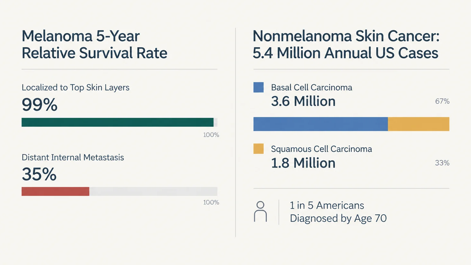
Clinical statistics emphasize why routine vigilance matters. Research shows that at least one in five Americans will develop skin cancer by age seventy.
When clinicians identify melanoma while it remains localized to the top skin layers, the five-year relative survival rate reaches ninety-nine percent. That figure drops to approximately thirty-five percent once abnormal cells metastasize to distant internal organs.
Nonmelanoma skin cancers represent the most prevalent malignancy worldwide. Over 5.4 million cases undergo medical treatment in the United States every single year.
Basal cell carcinoma accounts for roughly 3.6 million of these annual cases; squamous cell carcinoma accounts for another 1.8 million. Early excision preserves surrounding healthy tissue and prevents extensive functional damage.
Projections from the American Cancer Society indicate that over 100,000 new invasive melanoma cases are diagnosed annually. Rapid clinical identification remains the single most effective tool for preventing melanoma-related mortality.
Past ultraviolet exposure heavily dictates your individual risk profile. According to the Skin Cancer Foundation, experiencing five or more sunburns doubles your lifetime risk of developing melanoma.
A single blistering sunburn during childhood or adolescence also doubles your long-term risk. Ultraviolet-induced mutations accumulate quietly over decades before surfacing as visible lesions on your body.
Recognizing 7 Skin Spots Worth Getting Checked
1. The Asymmetrical, Evolving Mole
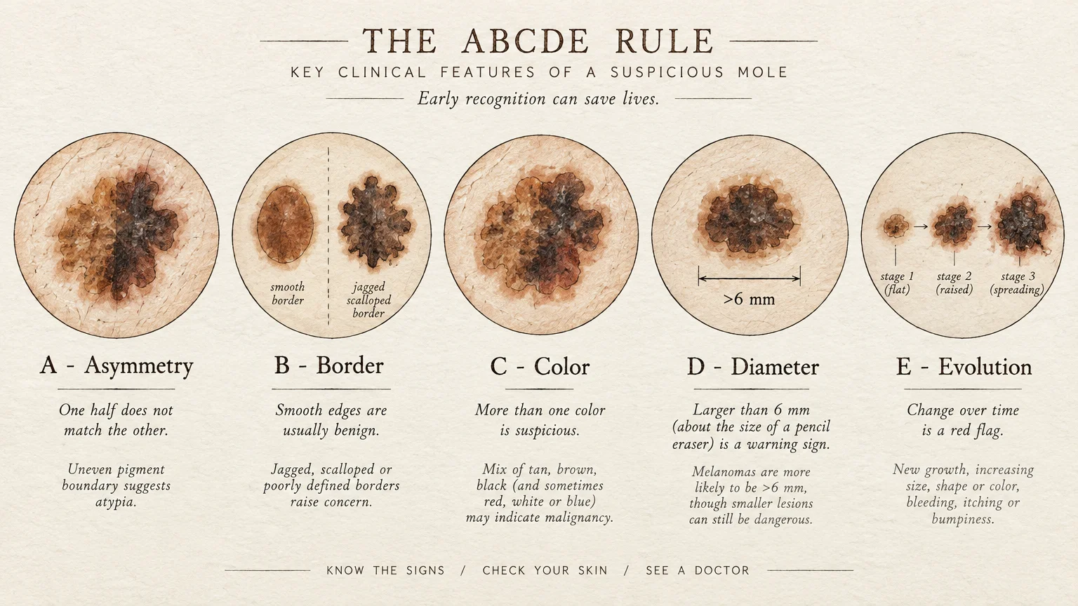
Dermatologists rely on the validated ABCDE criteria to diagnose melanoma in its earliest stages. This system tracks asymmetry, border irregularity, color variation, diameter, and evolution over time.
Benign moles usually present as symmetrical circles or ovals with smooth, defined borders. If you draw an imaginary line through your spot and the halves do not match, schedule an evaluation.
Standard diagnostic guidelines cite a diameter greater than six millimeters—about the size of a pencil eraser—as a primary warning flag. Yet early-stage melanomas can occasionally manifest as tiny, pinpoint dots.
The American Academy of Dermatology considers evolution the most critical parameter. Any existing mark that changes in size, shape, elevation, or shade warrants immediate professional scrutiny.
2. The Translucent, Non-Healing Sore
Basal cell carcinoma often disguises itself as an irritated patch or a persistent pimple that refuses to resolve. It commonly develops on sun-exposed zones like your nose, ears, neck, and shoulders.
These lesions frequently display a pearly, translucent quality and may feature slightly raised, rolled borders. You might notice minute blood vessels branching across the surface like delicate threads.
A classic warning sign involves a cycle of spontaneous bleeding, crusting over, and partial healing, followed by recurrence. True blemishes resolve within three to four weeks; malignant lesions linger indefinitely.
While basal cell carcinomas rarely metastasize to distant organs, they invade local cartilage, nerves, and underlying bone if neglected. Prompt removal prevents deep structural destruction.
3. The Rough, Sandpapery Patch
Actinic keratosis represents a widespread precancerous condition affecting roughly 58 million Americans. These spots often emerge as flat, rough, or scaly patches on chronically sun-damaged skin.
You will often feel an actinic keratosis before you clearly see it. Patients frequently describe the sensation as rubbing their fingertips against coarse sandpaper.
An individual actinic keratosis carries a five to ten percent risk of transforming into invasive squamous cell carcinoma over time. Furthermore, clinical data shows that over eighty percent of cutaneous squamous cell carcinomas arise directly from pre-existing actinic keratoses.
Dermatologists easily eliminate these precancerous cells using topical immunomodulators, field therapies, or liquid nitrogen cryotherapy. Treating them early halts potential malignancy before invasive growth begins.
4. The Firm, Rapidly Growing Crusted Nodule
Squamous cell carcinoma typically manifests as a firm, red nodule or an elevated, wart-like growth with a central depression. These growths frequently feel tender or sore when compressed.
With over 1.8 million cases diagnosed annually in the United States, squamous cell carcinoma ranks as the second most common form of skin cancer. It favors areas with substantial lifetime ultraviolet radiation, including your lower lip, scalp, and forearms.
These tumors may develop thick, stubborn crusts that bleed easily upon minimal trauma. Unlike superficial blemishes, they feel anchored into the deeper dermis.
Squamous cell carcinomas carry a distinct risk of spreading to regional lymph nodes if left untreated. Prompt surgical excision ensures complete removal and preserves your peace of mind.
5. The “Ugly Duckling” Lesion
Dermatologists rely on the ugly duckling sign to identify atypical lesions that escape standard diagnostic algorithms. Most people produce moles that resemble each other in color, size, and pattern.
An ugly duckling lesion stands out as an obvious visual outlier against your surrounding skin patterns. It may appear significantly darker, much paler, or considerably larger than your other marks.
This principle proves especially valuable if you possess dozens of atypical moles across your back and limbs. When one spot deviates noticeably from your body’s typical mole family, clinicians prioritize it for biopsy.
Trust your instincts when evaluating these visual outliers. If a spot simply looks odd or inconsistent compared to your other markings, request a dermatological evaluation.
6. Dark Streaks Beneath Nails or Pigment on Palms
Acral lentiginous melanoma develops on skin surfaces that receive little to no sunlight, such as your palms, soles, and nail beds. This rare subtype disproportionately impacts Black, Hispanic, and Asian individuals.
Because public health campaigns focus heavily on sun-exposed zones, patients frequently overlook these hidden areas. As a result, clinicians often diagnose acral melanoma at advanced, dangerous stages.
Look for a distinct vertical brown or black band running down your fingernail or toenail matrix. Warning signs include pigment that widens over time or spills over onto the surrounding cuticle skin.
You should also inspect your soles and palms for persistent, irregular dark patches that resemble stubborn bruises. A professional examination clarifies whether discoloration stems from physical trauma or cellular proliferation.
7. The Spreading, Mottled Patch: Age Spots vs. Melanoma
Differentiating harmless age spots from lentigo maligna melanoma represents a common clinical challenge. Age spots, known clinically as solar lentigines, emerge as smooth, uniformly tan or light brown marks.
Benign age spots typically maintain stable boundaries and consistent pigmentation over many years. They occur frequently on mature skin following cumulative lifetime sun exposure.
In contrast, lentigo maligna melanoma expands slowly across your skin surface over months or years. It presents with mottled, variegated shades of tan, dark brown, black, and sometimes red or white.
According to the National Cancer Institute, evolving pigmentation and jagged borders signal malignancy rather than normal aging. Any spreading pigmented patch demands careful dermoscopic assessment.

Clinical Insights and Patient Experiences
Practicing dermatologists emphasize that self-advocacy and early clinical contact consistently yield the best health outcomes. Clinical specialists observe that patients often wait months before showing an irregular spot to their physician.
“Patients often apologize for making an appointment over a spot that turns out benign,” explains Dr. Elena Vance, a clinical dermatologist. “We celebrate benign findings because catching the one malignant lesion early preserves a patient’s life.”
Patient stories highlight the lifesaving impact of rapid action. Marcus Reynolds, a marathon runner diagnosed with stage 1 melanoma at age thirty-four, credits regular self-exams with saving his life.
“I noticed a tiny dark spot on my calf that had not been there six months prior,” Marcus recalls. “My biopsy confirmed early melanoma, and a simple in-office excision cured it completely.”
Listening to both clinical guidance and personal intuition removes hesitation. Partnering with a skilled provider gives you clarity and actionable reassurance.

Pillars of Proactive Skin Health
Conducting Thorough Monthly Self-Examinations
Perform a head-to-toe skin self-examination once every month in a brightly lit room. Use a full-length mirror and a hand mirror to inspect hard-to-see areas like your scalp, back, and buttocks.
Part your hair using a comb or a blow dryer to inspect your scalp thoroughly. Examine your fingers, toes, nail beds, and the spaces between your digits.
Document suspicious marks by taking clear, well-lit digital photographs next to a coin or ruler for size reference. This visual baseline allows you and your clinician to identify subtle monthly changes accurately.
Practicing Daily Photoprotection
Preventing ongoing DNA damage requires consistent, comprehensive sun protection strategies. Guidelines from the Centers for Disease Control and Prevention recommend applying broad-spectrum sunscreen with an SPF of thirty or higher daily.
Reapply sunscreen every two hours during outdoor activities, or immediately after swimming or heavy sweating. Sun protection remains essential even on cloudy days because ultraviolet rays penetrate cloud cover effortlessly.
Complement topical sunscreens with physical shade, wide-brimmed hats, and ultraviolet protection factor clothing. Limiting midday sun exposure between ten in the morning and four in the afternoon dramatically lowers cumulative cellular stress.
Navigating Healthcare Access and Clinical Advocacy
Schedule an annual full-body examination with a board-certified dermatologist, especially if you have fair skin, family history, or numerous moles. These visits allow specialists to evaluate spots using high-powered epiluminescence microscopy.
If you face barriers accessing specialized dermatologic care, request a skin evaluation from your primary care physician. Many community health clinics and regional hospital networks offer free or low-cost annual screening events.
Bring a list of your specific questions and your photographed skin spots to your consultation. Proactive preparation ensures your clinician examines every lesion that causes you concern.
Frequently Asked Questions
How often should you get a professional skin cancer screening?
Most adults benefit from an annual full-body skin examination performed by a qualified dermatologist. Individuals with a personal history of skin cancer or multiple atypical moles may need evaluations every three to six months.
Consult your healthcare provider to determine an appropriate screening schedule based on your individual risk factors. Personalized timelines optimize early detection while minimizing unnecessary procedures.
Can skin cancer appear in places that never receive sun exposure?
Yes, skin cancers can emerge anywhere on your body, including your groin, soles, armpits, and inside your mouth. Melanomas in non-sun-exposed regions often link to genetic mutations rather than ultraviolet damage.
Thorough full-body checks must encompass these hidden areas to ensure complete coverage. Never ignore an atypical lesion simply because the location rarely sees daylight.
What happens during a professional dermatologist skin check?
Your dermatologist conducts a systematic visual inspection of your skin from your scalp down to your toes. They use a handheld illuminated microscope called a dermatoscope to examine deep pigment patterns.
If the clinician discovers a suspicious spot, they perform a brief, localized biopsy using local anesthesia. The tissue goes to a dermatopathologist for precise cellular analysis.
How do you distinguish an ordinary mole from a dangerous spot?
Normal moles remain stable, symmetrical, and uniform in color throughout adult life. In contrast, dangerous lesions evolve continuously, present ragged borders, or showcase multiple contrasting colors.
Whenever a spot tingles, bleeds, itches, or changes appearance, arrange an in-person clinical assessment. Only a trained medical provider can provide a definitive histological diagnosis.
Taking Your First Step Toward Lasting Peace of Mind
Protecting your skin health does not require overwhelming lifestyle overhauls; it begins with intentional, observant awareness. Dedicate ten minutes today to examine your skin and photograph any mark that causes you hesitation.
Ask yourself: when was the last time a professional examined your skin from head to toe? Scheduling an appointment bridges the gap between passive worry and empowered self-care.
Your body works tirelessly to protect you every day. Returning the favor with diligent attention and timely medical support ensures you thrive for decades to come.









Leave a Reply