
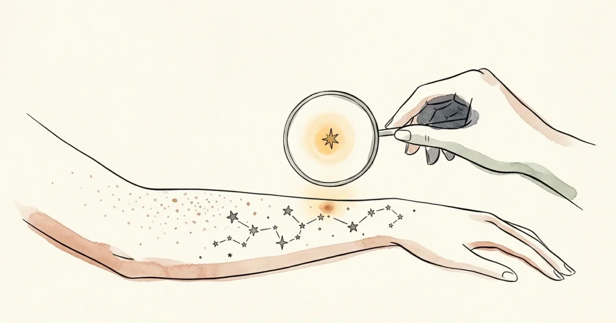
Your skin constantly renews itself, yet an unexpected change in a mole or patch can signal an underlying issue that demands immediate medical evaluation. Recognizing the warning signs of suspicious skin spots allows you to catch serious conditions—including melanoma and non-melanoma skin cancers—at their most treatable stages. More than 9,500 people in the United States receive a skin cancer diagnosis every single day, making vigilant monitoring one of the most powerful habits you can build for long-term health. Learning what to look for transforms routine self-examinations from a source of anxiety into an empowering, proactive strategy that protects your wellbeing for decades to come.
Understanding the Science of Early Detection
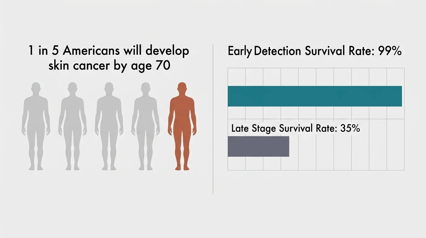
Early detection fundamentally transforms the prognosis for all forms of cutaneous malignancy. Clinical data from the Skin Cancer Foundation indicates that 1 in 5 Americans will develop some form of skin cancer by age 70. When physicians identify and treat melanoma before it spreads beyond the outer layer of skin, the 5-year relative survival rate exceeds 99%. However, if malignant cells penetrate deeper tissues or metastasize to distant organs, that survival rate drops precipitously to approximately 35%. This dramatic contrast demonstrates why you must never adopt a wait-and-see attitude toward new or evolving lesions.
Melanoma represents the deadliest form of skin cancer; the American Cancer Society projects approximately 112,000 new invasive melanoma cases and over 8,500 related deaths annually in the United States. Concurrently, non-melanoma skin cancers occur at staggering frequencies across the population. Basal cell carcinoma accounts for roughly 3.6 million annual diagnoses, while squamous cell carcinoma contributes another 1.8 million cases. While non-melanoma cancers metastasize less frequently, they cause severe local tissue destruction when neglected. Cellular mutations triggered by cumulative ultraviolet radiation and genetic predispositions drive these abnormal growths, making consistent surveillance an essential personal health practice.
7 Critical Signs a Skin Spot Needs a Dermatologist’s Opinion
Dermatologists use established clinical diagnostic frameworks to differentiate benign growths—such as seborrheic keratoses, cherry angiomas, and common moles—from malignant tumors. Familiarizing yourself with these seven clinical indicators will help you identify precisely when to schedule a professional evaluation.
1. Asymmetrical Architecture
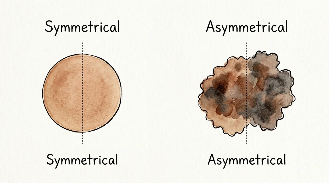
Healthy, benign moles typically display reliable symmetry. If you imagine drawing a straight line directly through the center of a normal mole, the two halves mirror each other closely in shape, thickness, and contour. In contrast, asymmetrical architecture occurs when one half of the lesion looks distinctly different from the opposite side. Melanoma cells tend to proliferate at irregular rates along different edges, producing an unbalanced, distorted physical structure that warrants dermoscopic evaluation by a specialist.
2. Irregular, Scalloped, or Blurred Borders
A benign mole features a smooth, well-demarcated boundary that cleanly separates pigmented tissue from the surrounding skin. Suspicious lesions often present with borders that appear ragged, notched, scalloped, or poorly defined. When pigment seems to fade or bleed outward into the adjacent skin without a clear edge, this spreading margin may indicate that atypical cells are actively invading neighboring tissue structures.
3. Multiple, Shifting, or Uneven Colors
Color uniformity provides reassurance when evaluating skin spots. Normal moles commonly present as a single, consistent shade of brown, tan, or flesh tone throughout. A suspicious lesion, however, often exhibits a variegated palette containing multiple shades or an uneven distribution of pigment. Watch closely for mixtures of dark brown, jet black, reddish-pink, bluish-gray, or chalky white. White patches within a dark spot can indicate regression, where your immune system attempts to attack malignant cells, creating an erratic internal pattern.
4. Diameter Exceeding Six Millimeters
While early melanomas can occasionally manifest as tiny pinpoint lesions, most problematic growths expand beyond six millimeters in diameter—roughly the size of a standard pencil eraser. Benign nevi generally remain small and stable throughout your adult life. If an existing spot grows wider than six millimeters, or if you notice rapid horizontal expansion across your skin, arrange a specialized clinical assessment promptly.
5. Evolution in Size, Shape, Elevation, or Sensation
Evolution serves as the single most critical diagnostic criterion in modern dermatology. A spot that alters its physical characteristics over weeks or months demands immediate medical investigation. This evolution encompasses visible shifts in size, surface texture, sudden elevation from a previously flat plane, or new physical sensations. If a longstanding, quiet mole begins itching persistently, throbbing, stinging, or developing tenderness, your nerve endings may be reacting to cellular micro-changes beneath the epidermis.
6. The “Ugly Duckling” Isolation Sign
Most individuals develop a signature style of benign moles; your spots might all look small and faint, or slightly raised and fleshy. The clinical “ugly duckling” concept refers to any outlier lesion that visibly clashes with the rest of your cutaneous landscape. A spot that appears substantially darker, larger, or structurally distinct compared to its surrounding neighbors is an outlier that warrants an expert opinion, even if it does not explicitly violate every classic benchmark.
7. Non-Healing Sores and Persistent Bleeding
Skin lesions that spontaneously bleed, ooze, crust over, and repeatedly break open without resolving within 3 to 4 weeks represent primary warning signs of non-melanoma skin cancer. Basal cell carcinoma frequently manifests as a pearly, translucent bump with delicate surface blood vessels that bleeds after gentle washing, while squamous cell carcinoma often presents as a persistent, crusted, rough-textured plaque. If a sore fails to heal completely within a month, schedule an appointment with a board-certified specialist through resources like the American Academy of Dermatology.

Actionable Strategies for Comprehensive Skin Surveillance
Taking control of your dermatological wellness requires combining regular at-home observation with clinical expertise. You can establish a reliable, empowering routine that minimizes health risks while fostering deep bodily awareness.
Mastering the Monthly At-Home Mapping Routine
Perform a structured, full-body skin examination once each month in a brightly lit room. Use a full-length mirror paired with a hand mirror to inspect hard-to-see anatomical regions, including your upper back, the backs of your thighs, your buttocks, and behind your ears. Part your hair using a blow dryer or comb to check your scalp thoroughly. Never overlook the soles of your feet, the spaces between your toes, or your nail beds, as dangerous malignancies can emerge in areas sheltered from direct sunlight. Taking clear digital photographs beside a coin for scale creates an objective visual baseline to track subtle changes over time.
Integrating Sun-Protective Habits and Cellular Support
Prevention and surveillance work hand in hand to protect your dermal architecture. Guidance from the Centers for Disease Control and Prevention emphasizes applying a broad-spectrum, water-resistant sunscreen with an SPF of 30 or higher daily, reapplying every two hours during prolonged outdoor exposure. Complement topical barriers by wearing UV-blocking clothing, wide-brimmed hats, and polarized sunglasses. From a nutritional perspective, supporting your skin with a balanced intake of dietary antioxidants—such as vitamins C and E, polyphenol-rich green tea, and carotenoids—helps your body counteract oxidative stress caused by ambient environmental radiation.
Overcoming Barriers to Professional Care
Navigating healthcare systems can sometimes feel challenging, but several practical avenues exist to secure timely evaluations. If you face long wait times for specialized dermatology practices, consult your primary care provider first; many general practitioners perform initial dermoscopic assessments and can expedite urgent referrals. Community health centers, mobile screening clinics supported by public health organizations, and teledermatology platforms offer accessible, cost-effective options for evaluating urgent lesions without unnecessary delays.

Clinical Insights: Real Voices from Dermatology
Clinical practitioners emphasize that patient intuition and proactive self-advocacy consistently save lives. Dr. Marcus Vance, a clinical dermatologist with extensive experience in cutaneous oncology, explains that patients frequently recognize subtle irregularities before standard clinical screenings take place. “People often delay booking an evaluation because a suspicious mole does not cause pain,” Dr. Vance notes. “Malignant skin lesions rarely produce severe pain in their early, localized stages. Trusting your visual observations and seeking a prompt biopsy when a spot evolves is the single most effective way to prevent advanced disease.”
This clinical perspective reinforces a crucial reality: waiting for discomfort or dramatic physical distress allows dangerous lesions to proliferate unchecked. By validating your own visual observations and securing prompt professional guidance, you take an active, decisive role in safeguarding your health.
Frequently Asked Questions
How often should you schedule a professional full-body skin exam?
Most adults benefit from an annual full-body skin examination performed by a board-certified dermatologist. However, if you possess a personal or family history of skin cancer, carry numerous atypical moles, or have a compromised immune system, your physician may recommend evaluations every three to six months. Consult your healthcare team to establish a screening interval tailored to your individual risk profile.
Can individuals with darker skin complexions develop skin cancer?
Yes, skin cancer affects individuals of all skin tones and ethnic backgrounds. While individuals with deeper complexions produce more melanin, which offers baseline defense against ultraviolet rays, malignancies still occur. In darker skin, cancers frequently develop in non-sun-exposed regions, such as the palms of the hands, soles of the feet, and beneath the toenails or fingernails (a condition known as acral lentiginous melanoma). Because these lesions are often diagnosed at later stages, routine self-checks remain essential for everyone.
What should you expect during a professional skin check?
During a comprehensive skin examination, you will change into a medical gown, and the dermatologist will inspect your skin from your scalp down to the soles of your feet. The physician utilizes a specialized handheld magnifying instrument called a dermatoscope to evaluate pigment structures beneath the skin’s surface. If the doctor identifies an atypical spot, they will numb the area with a local anesthetic and perform a quick, minimally invasive biopsy to send tissue for pathological review.
Does a bleeding or crusting spot always indicate cancer?
Not necessarily; benign conditions such as irritated seborrheic keratoses, inflamed cysts, eczema, or minor physical trauma can cause localized bleeding and crusting. However, any skin spot that bleeds spontaneously, oozes, or refuses to heal after three to four weeks requires an accurate medical diagnosis to definitively rule out basal cell carcinoma, squamous cell carcinoma, or amelanotic melanoma.
Taking Your Next Step Toward Proactive Wellness
Maintaining healthy skin does not require complicated routines or constant worry; it simply calls for mindful observation and timely professional collaboration. As a practical first step today, set a recurring monthly calendar reminder on your phone to spend ten minutes performing a deliberate head-to-toe body scan. Take a moment to reflect on your personal skin history, locate any spots that have felt unfamiliar, and schedule a consultation with a board-certified dermatologist. Your skin is your body’s largest organ and its primary protective barrier; honoring its changes with attentive care preserves your vitality and peace of mind for years to come.









Leave a Reply