

Your eyesight can change in an instant, turning an ordinary day into a critical medical emergency. Recognizing the early warning signs of a detached retina gives you the power to preserve your sight.
When delicate retinal tissue pulls away from underlying blood vessels, photoreceptor cells quickly starve of oxygen. Immediate clinical care stops this progressive damage before central vision disappears permanently.
Knowing what specific visual disturbances demand urgent attention helps you act with speed and confidence. Safeguarding your sensory connection to the world begins with understanding what your eyes are communicating.
Understanding the Mechanics of Retinal Detachment
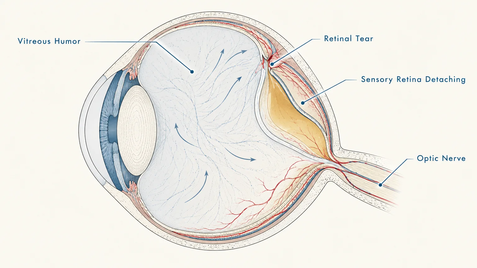
Your retina functions as the light-sensitive projection screen lining the interior back wall of your eye. It converts incoming light into electrical impulses that travel along the optic nerve to your brain.
A thick, clear gel called the vitreous humor fills the central cavity of your eye. As you age, this gel naturally shrinks, liquefies, and gently separates from the retinal surface.
Sometimes, this shrinking vitreous gel adheres too tightly and tears the fragile retinal tissue. Fluid then seeps through the resulting tear, peeling the sensory retina away from its underlying tissue bed.
Data from the National Eye Institute confirms that detached tissue loses its primary oxygen and nutrient supply. Starved photoreceptor cells quickly sustain irreversible cellular injury without prompt surgical intervention.
Rhegmatogenous retinal detachment represents the most common form of this condition. Eye specialists treat this mechanical detachment as a true eye emergency requiring urgent surgical evaluation.

8 Signs of a Detached Retina You Should Never Ignore
A detached retina causes no physical pain because the sensory retina lacks pain-sensing nerve fibers. You must rely entirely on distinctive visual changes to recognize when an eye emergency strikes.
1. A Sudden Shower of Floaters
Floaters appear as small specks, cobwebs, or translucent threads drifting across your field of view. While occasional floaters develop naturally with age, a sudden explosive outbreak serves as an urgent eye floaters warning.
This sudden shower occurs when tearing retinal vessels release microscopic red blood cells into your clear vitreous fluid. You might perceive this as looking through a dense swarm of gnats or a sprinkle of pepper.
2. Flashes of Light in Vision
Photopsia refers to brief flashes, sparks, or lightning streaks within your field of view. Experiencing sudden flashes of light in vision indicates that the shrinking vitreous gel is tugging mechanically against your retina.
Unlike migraine auras that shimmer and slowly fade over thirty minutes, retinal flashes are brief, sudden, and recurring. They appear most prominently in dim environments or when you rapidly shift your gaze.
3. A Dark Curtain Moving Across Your Field of View
A creeping dark curtain or shadow remains one of the most classic detached retina symptoms. Patients often report an opaque shade spreading inward from the outer periphery of their visual field.
This shadow corresponds directly to the anatomical area where the retina has peeled away from nourishing blood vessels. The curtain typically expands as fluid spreads, and resting will not make it fade away.
4. Rapid Loss of Peripheral Vision
Because retinal tears frequently originate in the far periphery of the eye, side vision often deteriorates first. You might bump into door frames, miss nearby objects, or feel like you are looking through a narrowing tunnel.
Central vision often stays deceptively clear during this early stage, leading people to postpone medical care. Regularly cover one eye at a time to verify that your full panoramic field remains crisp and unobstructed.
5. Blurry Vision That Arrives Without Warning
Sudden, unexplained blurriness in a single eye requires immediate diagnostic investigation by an eye specialist. When subretinal fluid spreads toward the macula, central visual sharpness degrades rapidly.
Blinking repeatedly, rubbing your eyelids, or applying lubricating eye drops will not resolve this internal haze. Prompt intervention before the macula fully detaches offers the best chance of preserving sharp reading vision.
6. Distorted or Wavy Lines
Metamorphopsia causes straight edges to look bent, irregular, or wavy. Looking at bathroom tiles, door frames, or lines of printed text will reveal these obvious spatial distortions.
Clinical guidance from the American Academy of Ophthalmology highlights this symptom as a critical warning. Distorted lines indicate that accumulated fluid has lifted and wrinkled your delicate macular tissue.
7. A Heavy or Hazy Gray Veil
Some individuals experience a dense gray or translucent film rather than a pitch-black shadow. This veil creates the frustrating impression of looking through frosted glass or cloudy water.
This effect occurs when mild vitreous bleeding blends with early localized retinal elevation. Colors lose their natural contrast and vibrancy, appearing noticeably washed out and muddy in the affected eye.
8. Sudden Blind Spots in Central Sight
The appearance of a solid central blind spot signals that the detachment has reached your fovea. This tiny central zone contains the dense cone photoreceptors responsible for sharp, detailed color sight.
Faces, books, and phone screens may disappear behind an empty gray or dark void. While emergency surgery can still reattach the tissue, treating tears before central detachment ensures vastly superior visual outcomes.
Clinical Perspectives on Emergency Intervention
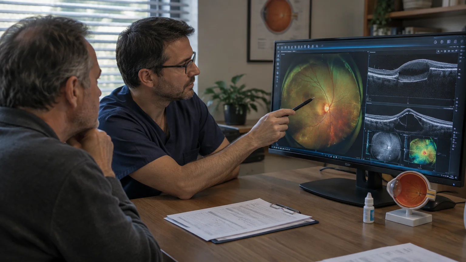
Ophthalmologists classify retinal detachments into two critical categories: macula-on and macula-off. A macula-on detachment represents an absolute medical emergency requiring surgical repair within twenty-four hours.
Retina specialists emphasize that repairing a detachment while the macula remains attached preserves reading vision in over ninety percent of cases. Once the macula separates, visual recovery becomes substantially less predictable.
Global health data from the World Health Organization highlights prompt vitreoretinal care as essential for preventing permanent blindness. Broadening public awareness of early retinal symptoms helps patients seek care before vision loss becomes irreversible.
Dr. Marcus Vance, a vitreoretinal surgeon, explains that time directly equates to preserved sight. He advises patients experiencing sudden floaters or flashes to bypass urgent care clinics and head straight to an ophthalmic hospital.
Strategic Pillars: Emergency Action, Recovery, and Prevention
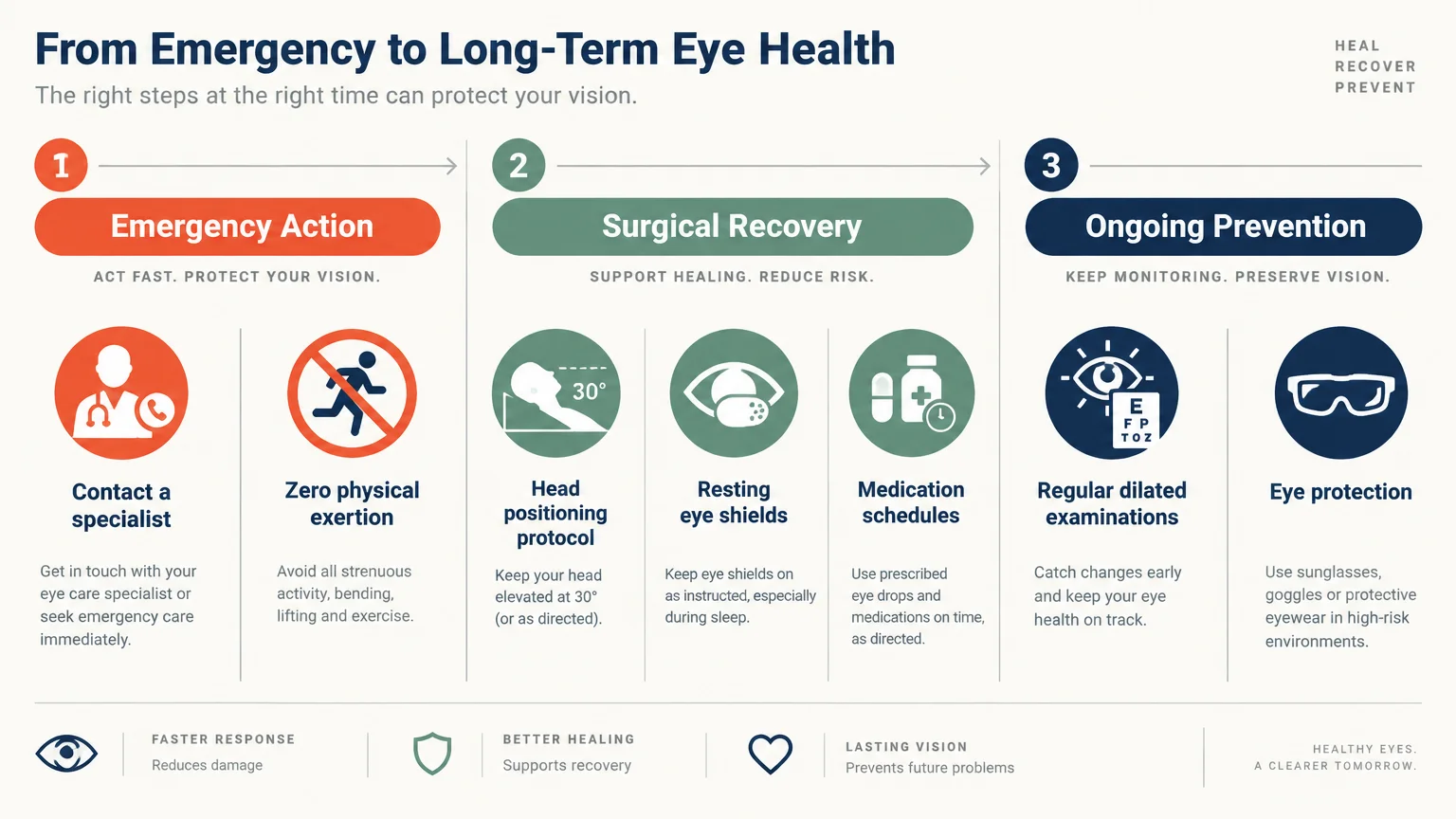
Safeguarding your eyesight requires efficient emergency navigation, rigorous post-procedure compliance, and proactive long-term wellness. These three strategic pillars empower you to protect your visual future effectively.
Pillar 1: Emergency Navigation and Medical Action
When you spot retinal detachment signs, navigating the healthcare system with speed saves your vision. Call a specialized ophthalmology practice or dedicated eye emergency room rather than booking a routine appointment.
Clearly state that you have sudden flashes, floaters, or a peripheral curtain over the phone. This clear terminology prompts medical triage teams to schedule you for an immediate same-day examination.
Your eye specialist will perform a comprehensive dilated fundus examination using an indirect ophthalmoscope. If caught early as a small tear, non-invasive laser photocoagulation or cryopexy can seal the tissue in the office.
Larger detachments require surgical reattachment through pneumatic retinopexy, scleral buckling, or a vitrectomy. Your surgeon will select the exact procedure that best secures your anatomical retinal alignment.
Pillar 2: Movement Safety and Post-Procedure Rest
Physical rest directly determines surgical success and proper retinal reattachment. If your surgeon injects an intraocular gas bubble during vitrectomy, maintaining strict head positioning becomes your primary duty.
You may need to maintain a strict face-down posture for several days after surgery. This precise alignment allows the buoyant gas bubble to float upward, pressing the torn retina against the ocular wall.
Avoid heavy lifting, intense workouts, and rapid head movements during your recovery window. These activities generate intraocular pressure spikes and physical shear forces that disrupt healing tissue layers.
Additionally, you must avoid flying or traveling to high elevations while a gas bubble remains in your eye. Reduced atmospheric pressure causes the intraocular bubble to expand dangerously, inducing severe secondary glaucoma.
Pillar 3: Mindset, Systemic Health, and Ocular Nutrition
Experiencing acute ocular symptoms triggers understandable fear and elevated stress levels. Practicing slow, diaphragmatic breathing activates your parasympathetic nervous system, stabilizing systemic blood pressure during recovery.
Your vascular health directly influences the microcirculation nourishing your retinal photoreceptors. Clinical research from the Centers for Disease Control and Prevention underscores managing blood pressure and glucose to preserve ocular microvessels.
Incorporate whole foods rich in lutein and zeaxanthin, such as cooked spinach, kale, and pasture-raised eggs. These dietary carotenoids accumulate within your macula, providing robust protection against oxidative stress.
Consume omega-3 fatty acids from wild salmon, flaxseeds, and walnuts to support retinal cell membrane flexibility. Maintaining steady daily hydration also supports normal vitreous gel integrity as you grow older.
Frequently Asked Questions
Does a detached retina cause physical eye pain?
No, a detached retina never causes physical eye pain, aching, or throbbing. The sensory retina does not possess pain-sensing nerve fibers.
Because physical discomfort is absent, you must rely entirely on visual warnings like flashes and floaters. Never delay seeking urgent medical evaluation simply because your eye feels comfortable.
Can a detached retina heal on its own without surgery?
No, a detached retina cannot heal spontaneously without medical treatment. Fluid will continue to seep beneath the retinal tear, widening the detachment over time.
Without surgical reattachment, chronic detachment leads to progressive, permanent blindness in the affected eye. Early surgical intervention by a vitreoretinal specialist is the only proven solution.
Who faces the highest risk for developing a detached retina?
People with high myopia, or severe nearsightedness, face significantly elevated risk because elongated eyeballs stretch retinal tissue. Individuals who have undergone cataract surgery or suffered eye trauma also carry greater vulnerability.
A personal or family history of retinal tears further increases your lifetime risk. Schedule yearly dilated eye exams to detect peripheral thinning before full tears emerge.
What should you do while traveling to an emergency eye clinic?
Keep your head steady and avoid rapid eye movements while traveling to the medical center. Do not read on your phone or bend over to pick up bags.
Have a family member or rideshare service drive you, as your vision may deteriorate quickly. Refrain from eating or drinking in case your ophthalmologist schedules immediate same-day surgery.
Preserving Your Vision for Life
Your eyesight is an irreplaceable gift that connects you to your loved ones and daily joys. Treating sudden visual symptoms with prompt respect is an essential act of self-care.
Take a practical first step today by finding and saving the contact details for your nearest retinal specialist. Having this emergency information readily available gives you invaluable peace of mind.
Take a brief moment right now to cover each eye separately and check your panoramic vision. Are you ready to prioritize and protect your eyesight through active, lifelong awareness?








Leave a Reply