

Recognizing subtle shifts in your eyesight protects your independence and shields you from permanent vision loss. Cataracts and glaucoma develop quietly, yet catching their warning signs early gives you the power to preserve your sight.
While both conditions alter how light reaches your brain, their biological mechanisms require completely different medical interventions. Understanding these distinct warning signs helps you seek targeted care before irreversible optic nerve damage or profound lens clouding occurs.
This clinical guide outlines ten critical vision changes you must never ignore. Learn what causes them and discover the exact steps you can take today to protect your long-term eye health.
Understanding the Difference Between Cataracts and Glaucoma
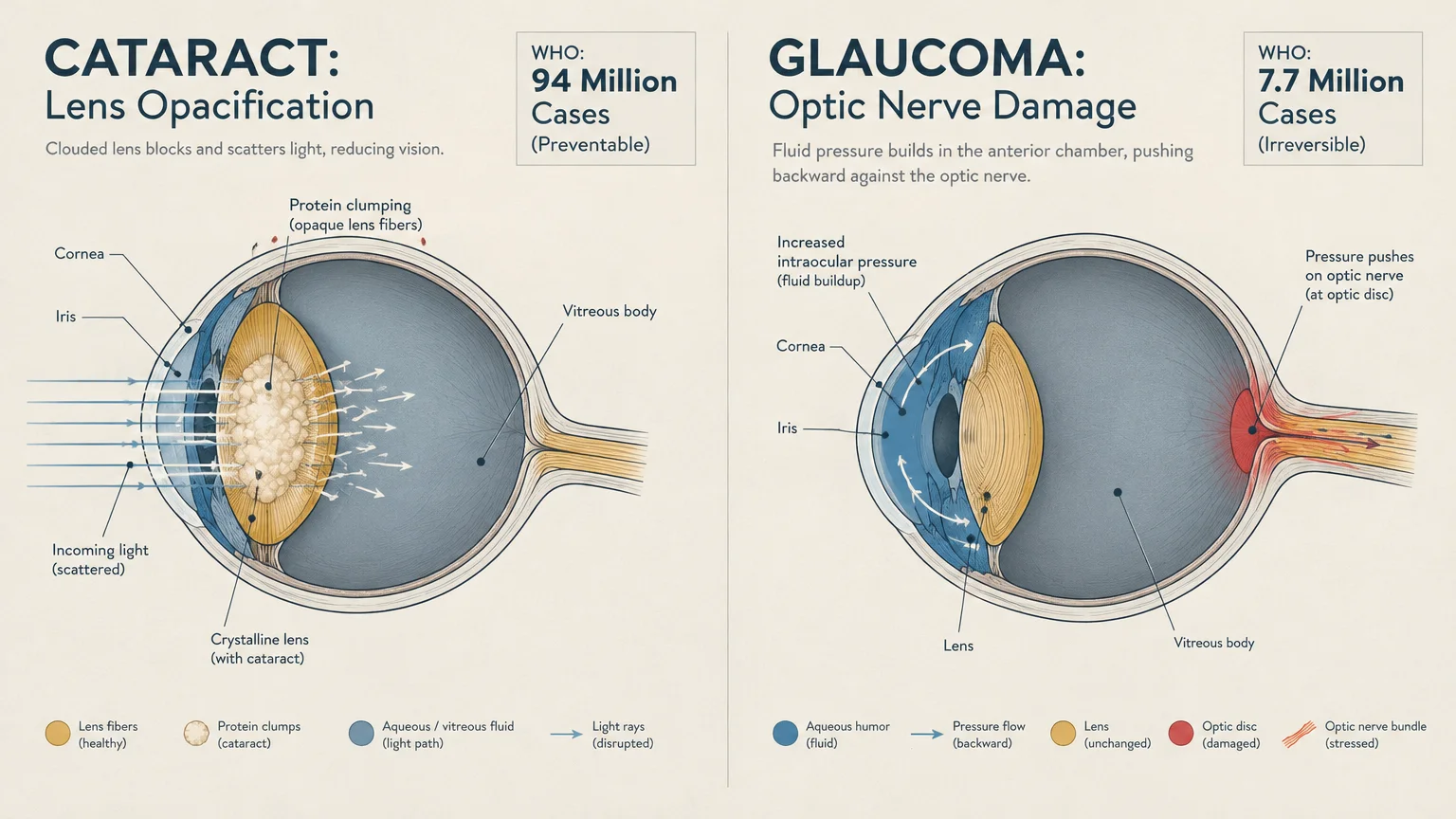
Cataracts occur when the naturally clear crystalline lens inside your eye becomes clouded by clumping structural proteins. This opacification scatters light, preventing sharp images from focusing directly onto your retina.
Glaucoma involves progressive damage to your optic nerve, which transmits visual electrical signals from your retina to your brain. This damage is frequently driven by an accumulation of intraocular fluid pressure.
According to the World Health Organization, cataracts account for 94 million cases of preventable distance vision impairment globally. This condition remains the leading cause of blindness worldwide.
The World Health Organization also identifies glaucoma as the leading cause of irreversible blindness across the globe, affecting 7.7 million people with unaddressed vision loss.
On a broader scale, approximately 80 million people worldwide live with glaucoma today. Open-angle glaucoma accounts for about 60 million of those global cases.
In the United States, surveillance estimates published by the Centers for Disease Control and Prevention indicate that 4.22 million adults live with glaucoma. This includes approximately 1.49 million Americans who experience vision-affecting glaucoma.
Cataracts represent an almost universal aspect of physical aging. The National Eye Institute notes that more than half of all Americans aged 80 or older either have cataracts or have undergone cataract surgery.
Distinguishing between these two conditions is essential for protecting your sight. While surgeons can safely replace a clouded cataract lens, medicine cannot restore optic nerve tissue once glaucoma destroys it.
10 Warning Signs of Cataracts and Glaucoma
Your eyes frequently provide subtle warnings before significant visual disability develops. Recognizing these ten signs allows you to consult an eye care provider before preventable or irreversible damage occurs.
1. Cloudy, Misty, or Blurred Central Vision
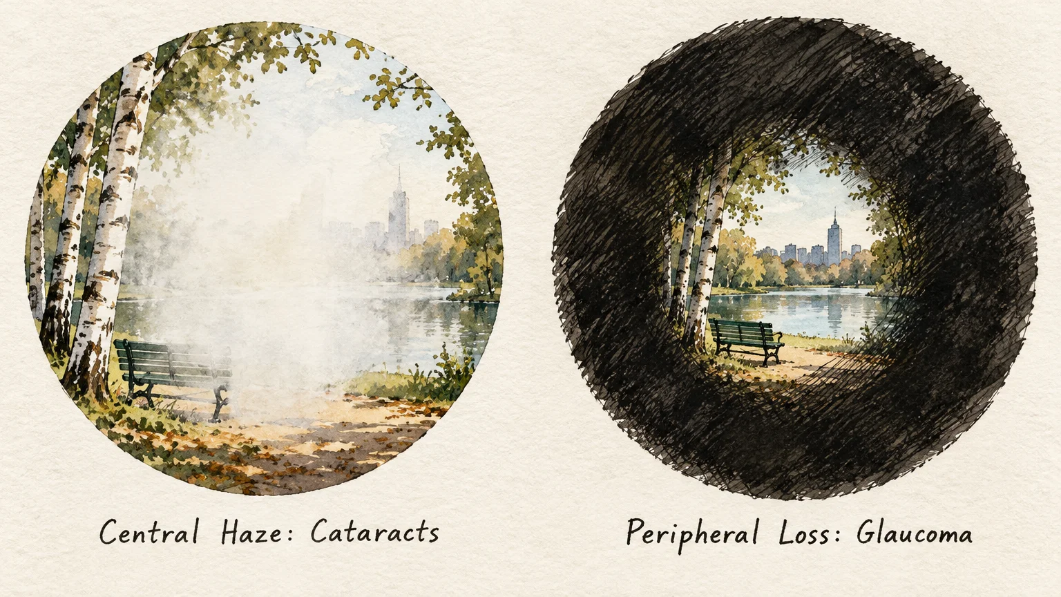
A classic symptom of cataract development is blurry or hazy central vision that resembles looking through a foggy window. You might wipe your spectacles repeatedly, only to realize the haze persists internally.
This haziness occurs because structural proteins within your crystalline lens break down and coagulate. As these protein clumps expand, they block light rays from focusing precisely onto your retina.
Unlike temporary dryness, this optical cloudiness does not resolve when you blink or apply artificial tears. It gradually worsens over months or years, dimming fine details during reading and close work.
Glaucoma rarely causes this direct central haze in its early phases. If your vision feels perpetually cloudy across your primary line of sight, cataracts are the primary suspect.
2. Gradual Loss of Peripheral Vision
Open-angle glaucoma typically attacks your peripheral visual field first, eroding the edges of what you see. Because this decline happens incrementally, you may not notice the subtle shrinking of your visual boundaries.
Your brain naturally compensates for missing visual inputs by blending surrounding visual patterns. As a result, many people adapt unconsciously until significant optic nerve fibers have already perished.
You might notice this deficit when you bump into low furniture or fail to notice people walking beside you. In advanced stages, this peripheral erosion creates pronounced tunnel vision.
If you notice yourself turning your head frequently to locate items that were once easily visible, request an automated visual field test.
3. Glare and Halos Around Lights
Seeing glowing rings, halos, or starbursts around light sources is a common warning sign for both conditions. Oncoming car headlights or streetlamps may appear surrounded by intense, distracting glare during nighttime driving.
In cataracts, light diffraction causes this visual distortion. The dense, irregular protein clusters inside your clouded lens scatter incoming photons across your eye rather than focusing them cleanly.
In glaucoma, halos often signal an acute elevation in intraocular pressure. Rapid pressure spikes cause fluid to accumulate inside your cornea, creating microscopic corneal edema that breaks light into rainbow-colored circles.
If you observe sudden iridescent halos paired with ocular ache, seek emergency ophthalmic care immediately.
4. Fading, Yellowing, or Browning of Colors
As a cataract matures, your natural crystalline lens gradually hardens and turns a yellowish or brownish shade. This internal discoloration acts like an amber camera filter over your vision.
Rich hues like blues, purples, and greens begin to look washed out and dull. You may find it difficult to tell dark blue socks from black ones when dressing in the morning.
Because this tinting progresses slowly over several years, your brain gradually accepts the muted palette as normal. Most patients only realize how much color vibrancy they lost after cataract surgery restores clear optics.
Glaucoma can also impair subtle color discrimination in its later stages, but widespread color yellowing points squarely toward lens opacification.
5. Frequent Prescription Changes and “Second Sight”
Needing updated prescription eyeglasses every few months suggests that your eye lens is undergoing structural shifts. Cataract formation alters the thickness and curvature of your lens, constantly shifting your refractive error.
In some cases, adults with developing nuclear cataracts experience a curious temporary phenomenon known as second sight. Your close-up reading vision may unexpectedly improve, allowing you to read books without magnifying glasses.
This apparent improvement occurs because the center of your lens swells, temporarily making your eye more nearsighted. However, this transient benefit soon gives way to increased cloudiness and severe visual degradation.
Never ignore rapid refractive fluctuations; discuss them promptly with your optometrist to determine whether lens hardening is the driving factor.
6. Severe Eye Pain, Nausea, and Sudden Redness
While open-angle glaucoma develops painlessly, acute angle-closure glaucoma presents as a medical crisis. This emergency arises when your iris bulges forward, suddenly blocking the drainage angle where aqueous humor exits your eye.
This blockage triggers a dangerous surge in intraocular pressure within minutes or hours. You will experience intense eye pain, severe frontal headaches, eye redness, nausea, and persistent vomiting.
Acute angle-closure glaucoma can cause irreversible blindness within twenty-four to forty-eight hours if left untreated. Clinicians must administer emergency medications or perform immediate laser iridotomy to restore fluid drainage.
If you experience severe orbital aching alongside blurred vision and gastrointestinal distress, proceed to an emergency department without delay.
7. Monocular Double Vision (Diplopia)
Double vision that persists when you cover one eye is known clinically as monocular diplopia. This specific visual distortion points directly to a localized problem within your eye’s crystalline lens or cornea.
In cataract patients, uneven protein densities split incoming light beams into multiple pathways before they reach the retina. This creates ghost images or secondary shadows around letters, road signs, and objects.
Neurological double vision disappears when you close either eye, whereas cataract-induced double vision remains visible through the affected eye.
An ophthalmologist can quickly determine whether an irregular lens opacification causes your ghost images during a slit-lamp examination.
8. Declining Night Vision and Loss of Contrast
Finding it difficult to navigate in dim environments or drive safely after sunset is a prominent red flag. Both cataracts and glaucoma significantly impair your eye’s contrast sensitivity.
Cataracts physically block photons from penetrating your eye, which makes dimly illuminated rooms feel oppressively dark. You might notice yourself requiring much brighter lamps to read or prepare meals comfortably.
Glaucoma damages retinal ganglion cells that process low-light contrast and motion detection. As these cells deteriorate, distinguishing curbs from sidewalks or detecting dark obstacles at dusk becomes hazardous.
If night driving creates intense anxiety or disorientation, schedule a comprehensive visual exam to assess your optical clarity and optic nerve health.
9. Patchy Blind Spots in Your Visual Field
In open-angle glaucoma, microscopic damage to your optic nerve fibers creates localized dead zones called scotomas. These patchy blind spots often begin in your paracentral or nasal field of view.
Because your eyes share overlapping fields of vision, your brain skillfully pastes surrounding colors over these missing patches. You might only notice a blind spot when an object suddenly appears out of nowhere.
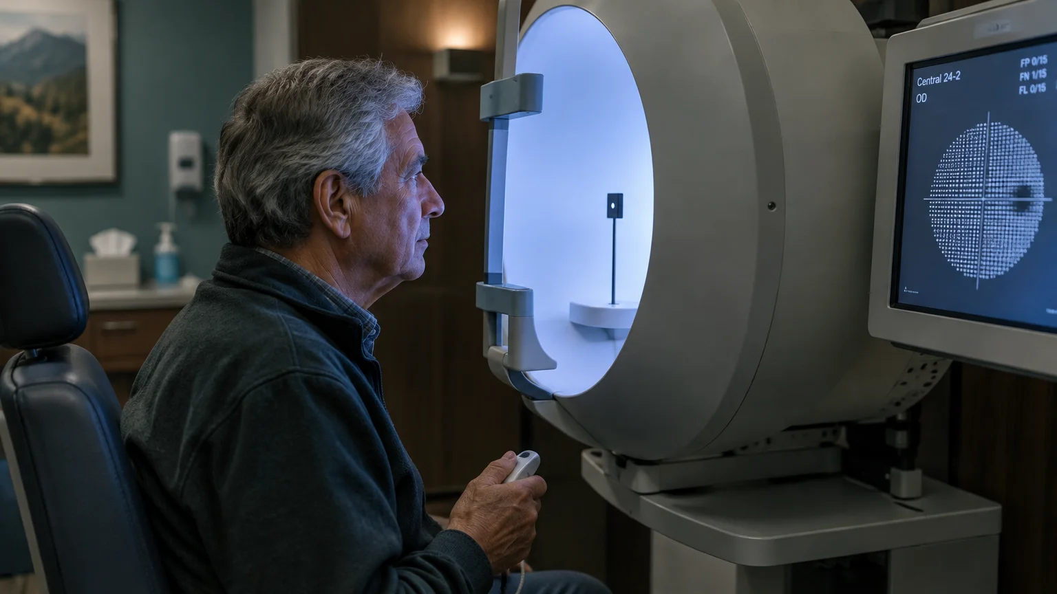
As the disease advances, these isolated patches coalesce into broad blind regions. Advanced automated perimetry at your eye clinic can map these invisible scotomas long before you consciously detect them.
Identifying scotomas early gives your clinical team the opportunity to lower eye pressure and protect your remaining functional vision.
10. The Complete Absence of Early Warning Signs
Perhaps the most perilous sign of glaucoma is the total absence of early symptoms. For this reason, ophthalmologists routinely call open-angle glaucoma the silent thief of sight.
CDC data confirms that approximately 50% of people living with glaucoma do not know they have it. Elevated fluid pressure does not cause itching, redness, soreness, or noticeable early vision loss.
By the time you notice physical gaps in your vision, significant and irreversible optic nerve damage has already occurred. You cannot rely on physical discomfort or overt blurriness to alert you to danger.
Proactive, scheduled clinical screenings remain your only dependable line of defense against asymptomatic optic nerve attrition.
Science Snapshot: Mechanisms and Risk Factors Behind Disease Progression
Clinical research demonstrates that both mechanical strain and metabolic oxidative stress drive visual decline as your eyes age. Understanding these pathways empowers you to make informed decisions regarding preventive care.
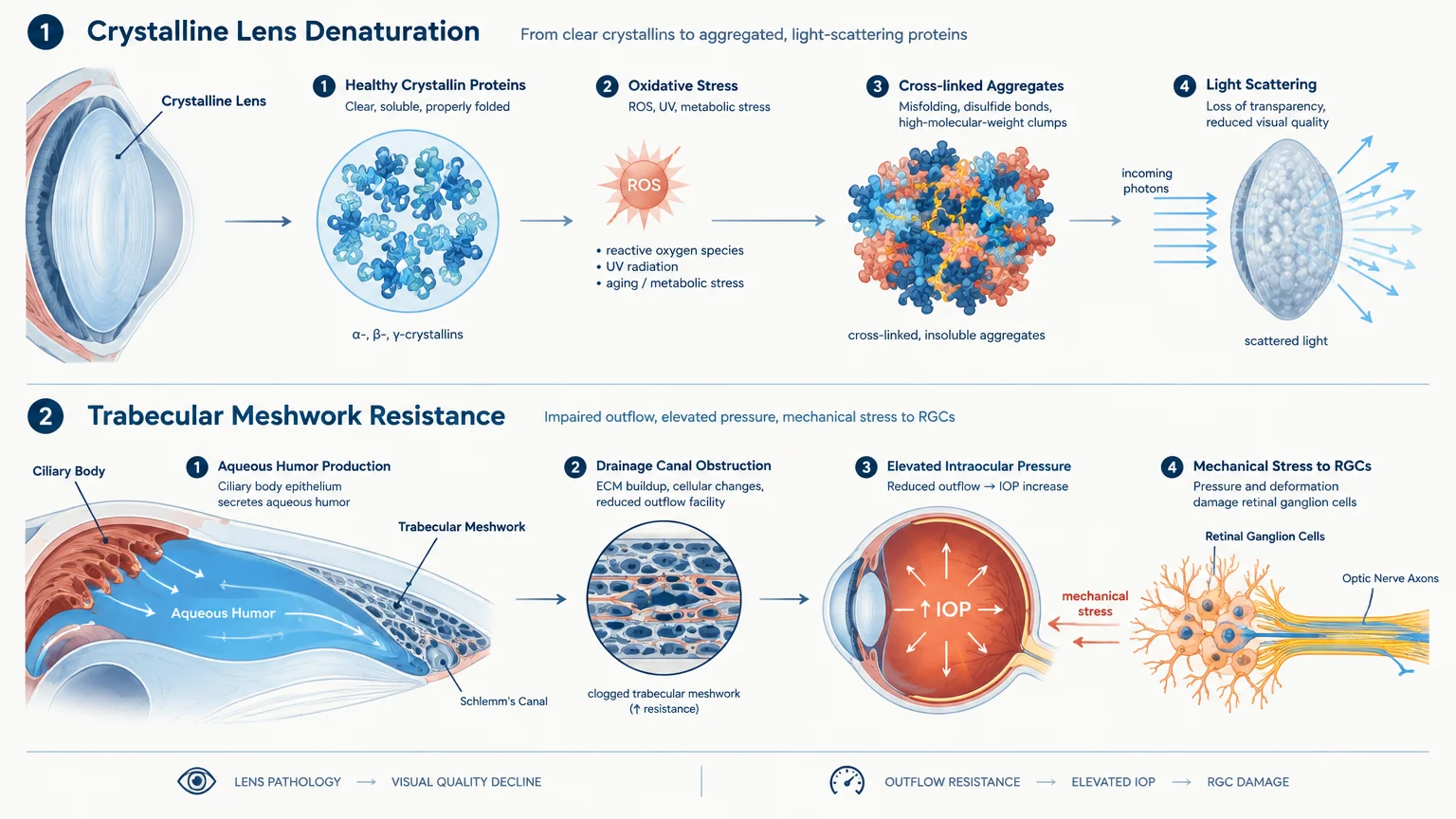
Glaucoma centers on the lamina cribrosa, a flexible sieve-like plate at the back of your eye. When intraocular pressure rises above what your optic nerve can tolerate, this plate bows backward.
This mechanical displacement pinches the delicate retinal ganglion cell axons running through the plate pores. The resulting disruption blocks axonal transport, cutting off crucial neurotrophic factors and triggering programmed cell death.
Epidemiological surveillance reveals severe demographic disparities in glaucoma vulnerability across populations. In the United States, Black adults have the highest prevalence rate among adults 18 and older at 3.15%.
By comparison, White adults demonstrate a prevalence rate of 1.42%. Furthermore, glaucoma-related blindness occurs 6 to 8 times more frequently in African Americans than in Caucasians.
Genetic variations in optic nerve structure, thinner central corneas, and microvascular differences contribute heavily to this increased risk profile. Knowing your heritage helps you and your physician determine an appropriate screening schedule.
Cataract formation, by contrast, represents cumulative oxidative damage within the structural proteins of your lens. Your natural lens cannot shed older cells; instead, it compresses older fibers into the center of the lens capsule.
Over decades, ultraviolet radiation, metabolic byproducts, and systemic inflammation cause these crystalline proteins to cross-link and denature. This biochemical change gradually turns a clear living optical lens into a cloudy, rigid barrier.

Actionable Strategy Pillars for Long-Term Eye Health
Preserving your vision over a lifetime requires a systematic framework that blends professional medical diagnostics with targeted daily habits. Focus on these four pillars to protect your optical system.
Pillar 1: Rigorous Medical Surveillance and Diagnostic Testing
You cannot monitor intraocular pressure or detect microscopic lens changes through self-examination at home. A comprehensive dilated examination by an optometrist or ophthalmologist provides an unhindered view of your internal ocular anatomy.
Ensure your eye doctor performs applanation tonometry to measure your fluid pressure and dilated ophthalmoscopy to inspect your optic cup. These baseline checks reveal early structural abnormalities before functional vision loss begins.
Request optical coherence tomography (OCT) imaging during your visits. OCT utilizes light waves to capture cross-sectional images of your retina, measuring your retinal nerve fiber layer thickness down to the micrometer.
Ask your provider to assess your central corneal thickness using ultrasound pachymetry. Thinner corneas yield artificially low pressure readings during standard tonometry, which can mask dangerous intraocular pressure spikes.
Follow the clinical screening intervals published by the American Academy of Ophthalmology. The AAO recommends that all adults receive a comprehensive baseline eye disease screening at age 40.
Between ages 40 and 54, schedule an eye examination every 2 to 4 years. For individuals aged 55 to 64, the AAO recommends an examination every 1 to 3 years.
At age 65 and older, you should see an eye doctor every 1 to 2 years. The American Optometric Association recommends annual examinations for all adults older than 60.
Pillar 2: Nutrition, Hydration, and Vascular Support
Your eyes possess some of the highest metabolic demands and densest capillary networks in your entire body. Nourishing these tissues with targeted micronutrients enhances cellular resilience against oxidative stress.
Prioritize dietary carotenoids by consuming dark leafy greens like spinach, kale, and collards alongside pasture-raised egg yolks. Lutein and zeaxanthin accumulate directly inside your crystalline lens, neutralizing free radicals and filtering blue light.
Support vascular endothelial function by incorporating cold-water fatty fish, chia seeds, and walnuts into your weekly meals. Omega-3 fatty acids encourage healthy microcirculation and help sustain appropriate ocular surface moisture.
Maintain stable blood glucose and blood pressure levels through balanced, whole-food nutrition. Chronic hyperglycemia accelerates cataract maturation through advanced glycation end-products, while uncontrolled hypertension damages the microvessels nourishing your optic nerve.
Avoid extreme nocturnal hypotension, especially if you take systemic antihypertensive medications before bedtime. Overly suppressed nighttime blood pressure starves your optic nerve of vital perfusion, potentially accelerating normal-tension glaucoma.
Stay properly hydrated throughout the day by sipping water consistently rather than chugging large volumes at once. Rapidly consuming a liter of water can trigger temporary spikes in intraocular fluid pressure.
Pillar 3: Protective Movement, Ergonomics, and Physical Habits
Regular physical movement supports healthy ocular perfusion and helps lower baseline intraocular pressure. Engaging in moderate aerobic exercise—such as brisk walking, cycling, or swimming—enhances neurovascular health throughout your visual pathway.
If your clinician has diagnosed you with glaucoma or ocular hypertension, avoid prolonged inverted body positions. Headstands, inversion therapy tables, and extended yoga poses like downward-facing dog significantly increase intraocular pressure.
Avoid heavy weightlifting exercises that require the Valsalva maneuver, such as holding your breath while straining against heavy resistance. Exhaling steadily during exertion prevents transient, sharp pressure elevations within your thoracic cavity and eyes.
Shield your eyes from solar ultraviolet radiation whenever you step outside during daylight hours. Choose sunglasses labeled UV400 or offering 100% UVA and UVB protection to prevent accelerated cataractogenesis.
Wear ANSI-rated safety goggles whenever you perform home maintenance, woodworking, gardening, or high-impact athletic activities. Direct ocular blunt trauma can damage your trabecular meshwork, provoking secondary angle glaucoma years later.
Pillar 4: Environmental Adaptations and Stress Management
Adapting your physical living space reduces visual fatigue and minimizes fall hazards as your eyes undergo natural age-related shifts. Simple environmental modifications provide immediate functional security.
Install adjustable, glare-free task lighting over reading nooks, kitchen worktops, and work desks. Position lamps so light shines directly onto your tasks without reflecting harshly into your eyes.
Create strong visual contrast along architectural edges to improve spatial navigation throughout your home. For example, use contrasting tape or distinct paint colors on the edges of stair treads and doorways.
Practice visual pacing when working on digital screens by observing the 20-20-20 protocol. Every 20 minutes, shift your focus to an object 20 feet away for 20 seconds to relax your focusing muscles.
Integrate intentional stress-reduction practices like slow diaphragmatic breathing and mindfulness into your daily schedule. Chronic psychological distress elevates systemic cortisol and sympathetic tone, which can adversely affect ocular hemodynamics.
Clinical Perspectives: What Specialists and Patients Experience
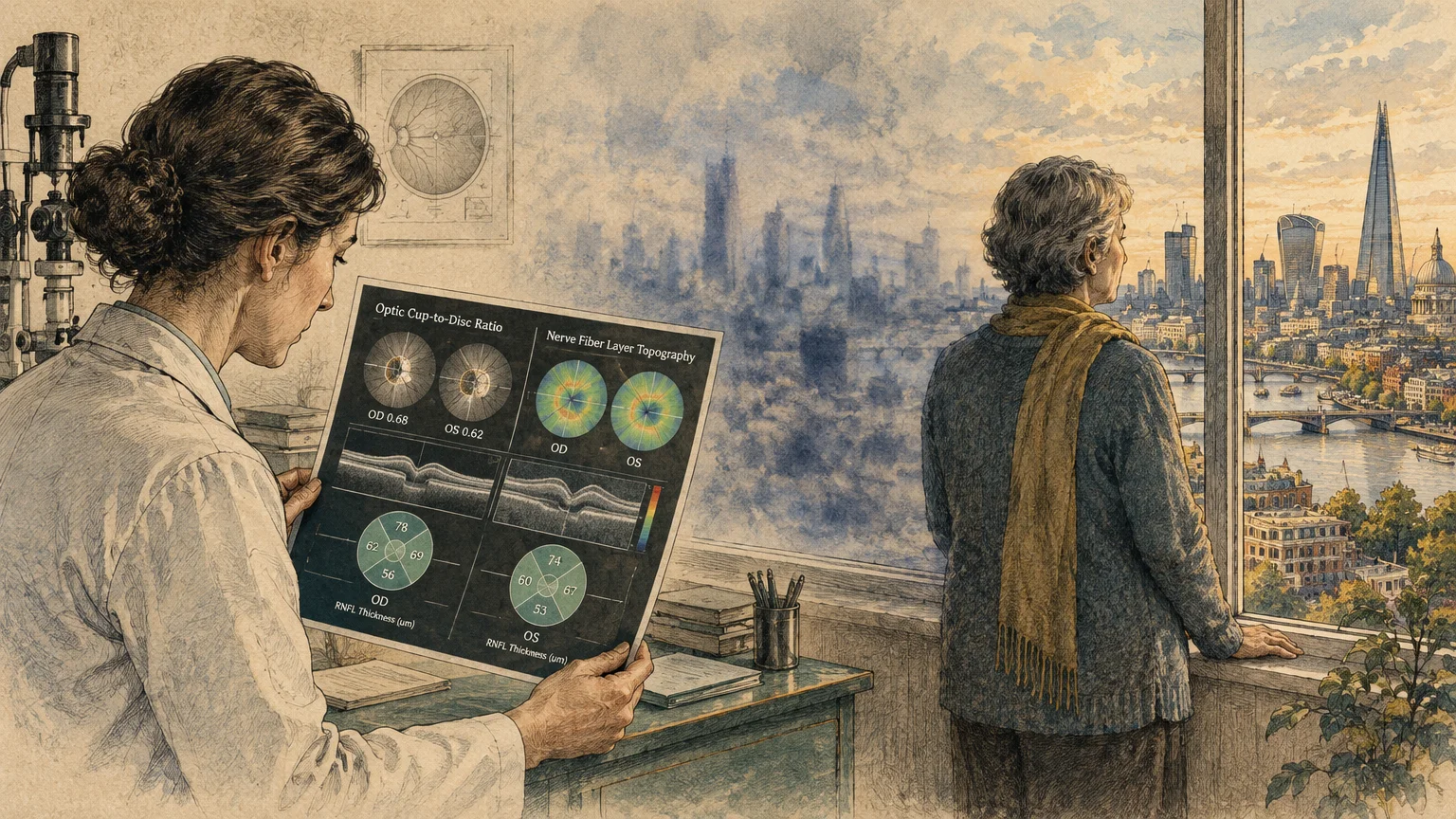
Clinicians repeatedly observe that patients delay scheduling eye examinations because they mistake progressive vision loss for normal, harmless aging. Experienced eye doctors stress that significant visual impairment is never an inevitable part of growing older.
Ophthalmologists note that individuals often adapt unconsciously to worsening cataracts by abandoning reading, driving, or creative hobbies. When these patients finally undergo cataract surgery, they frequently marvel at how much color and visual independence they surrendered.
Glaucoma specialists emphasize that consistency with prescribed pressure-lowering eye drops is the single most important factor in preserving sight. Missing daily doses allows intraocular pressure to fluctuate, quietly accelerating irreversible optic nerve atrophy.
Patient support organizations highlight the emotional relief that comes from obtaining an early, definitive diagnosis. Knowing your clinical status empowers you to collaborate with your medical team and implement therapies before your lifestyle suffers.
Open communication between you and your eye care specialist ensures that medication side effects—such as surface dryness or redness—are resolved promptly through alternative therapies.
When to See an Eye Doctor: Official Clinical Guidelines
Understanding clinical screening benchmarks prevents you from falling through diagnostic gaps. Both the American Academy of Ophthalmology and the American Optometric Association establish clear timelines based on age and clinical risk.
Schedule your comprehensive baseline eye disease screening at age 40, even if your vision feels entirely crisp and trouble-free. This evaluation provides your clinician with baseline measurements of your optic nerve anatomy and corneal thickness.
Between ages 40 and 54, maintain an exam schedule of every 2 to 4 years if you lack risk factors. Between ages 55 and 64, increase your visits to every 1 to 3 years.
Once you reach age 65, the AAO recommends an evaluation every 1 to 2 years. The AOA recommends that all adults aged 61 and older undergo annual comprehensive eye examinations.
You must schedule more frequent screenings if you have diabetes, systemic hypertension, a direct family history of glaucoma, or African ancestry. These clinical factors markedly increase your probability of developing optic nerve or vascular complications.
If you experience sudden visual field shadows, severe eye pain, or flashes of light, seek immediate same-day ophthalmic assessment.
Frequently Asked Questions About Cataracts and Glaucoma
Can lifestyle changes reverse cataracts or glaucoma?
No diet, nutritional supplement, or physical exercise can dissolve an existing cataract or regenerate damaged optic nerve fibers. Once crystalline proteins clump or retinal ganglion cells die, those physical changes remain permanent.
Healthy lifestyle habits do, however, slow down disease progression and protect your remaining functional vision. You should use evidence-based lifestyle strategies as supportive companions to formal medical and surgical care.
How do I distinguish between normal age-related vision changes and disease?
Normal aging commonly leads to presbyopia, a benign loss of near-focusing flexibility caused by the natural stiffening of your lens. Presbyopia makes reading small print difficult, but standard reading glasses immediately resolve the issue.
Eye diseases like cataracts and glaucoma produce symptoms that reading glasses cannot correct, including patchy blind spots, constant blur, and glare. Any visual distortion that resists corrective lenses requires a comprehensive diagnostic evaluation.
What happens during cataract surgery and glaucoma treatment?
Cataract surgery is a brief, highly reliable outpatient procedure that replaces your clouded natural lens with a clear intraocular lens implant. The surgeon removes the opacified lens using gentle ultrasonic vibration before positioning the new lens.
Glaucoma therapy focuses on reducing intraocular fluid pressure through daily prescription eye drops, in-office laser trabeculoplasty, or microsurgical filtration procedures. While these treatments cannot restore lost optic nerve tissue, they halt further visual field loss.
Can I have normal eye pressure and still develop glaucoma?
Yes, you can develop normal-tension glaucoma even if your intraocular pressure stays within conventional statistical limits. In this condition, the optic nerve sustains progressive damage despite pressure measurements falling under 21 mmHg.
This vulnerability often stems from vascular dysregulation, structural fragility at the lamina cribrosa, or reduced optic nerve perfusion. Consequently, measuring eye pressure alone is never enough to rule out glaucomatous optic neuropathy.
Your Next Step Toward Lifelong Vision
Taking decisive control of your visual wellness starts with a single concrete action today. Do not wait for noticeable blind spots, severe glare, or misty vision to compromise your daily independence.
Check your calendar and determine the date of your last comprehensive dilated eye exam. If you are past due based on current clinical guidelines, call an eye care specialist and schedule your evaluation today.
Reflect on how your vision has performed during daily tasks over the past few weeks. Have you experienced increased glare while driving, or needed brighter lighting to read comfortable text?
Note these subtle observations on paper and share them openly with your doctor during your visit. Being proactive ensures you protect your visual clarity and continue enjoying the activities and people you cherish.








Leave a Reply