
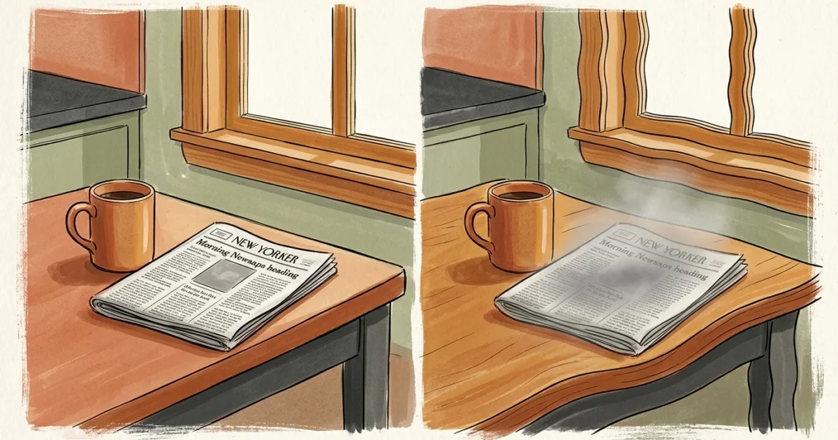
Assuming that worsening eyesight is merely an inevitable tax on aging often causes people to miss early, treatable warnings of underlying ocular disease. While mild difficulty with close-up reading naturally develops over time, severe visual changes—such as sudden distortion, dark spots, or loss of side vision—signal significant health shifts that demand immediate attention. Proactively monitoring your eye health allows you to safeguard your independence, maintain your quality of life, and protect your precious sight for decades to come. Recognizing the subtle distinction between normal physical maturation and pathology empowers you to seek professional care before permanent damage occurs.

Understanding Normal Aging Versus Pathological Vision Changes
As your body matures, your eye lenses gradually lose flexibility. This natural process, known as presbyopia, usually begins in your early-to-mid 40s and makes reading fine print or focusing on nearby objects challenging. You might find yourself holding menus at arm’s length or adjusting ambient light to read comfortable text; these adjustments represent predictable, benign shifts in visual focus.
Pathological vision changes operate through entirely different structural mechanisms. Rather than simple lens stiffness, serious ocular diseases damage the optic nerve fibers, light-sensing retinal cells, or delicate ocular blood vessels. Recognizing when a visual shift transcends ordinary presbyopia helps you seek medical evaluation quickly, protecting your sight against irreversible structural harm.

7 Vision Changes You Should Never Ignore
1. Cloudiness or Blurred Central Visual Fields
When everyday tasks like reading fine print or recognizing familiar faces begin to feel like looking through a foggy window, you may be experiencing signs of cataracts. Unlike temporary eye fatigue, cataracts occur when natural proteins inside your crystalline lens break down and clump together over time. This nuclear or cortical clouding blocks light rays from focusing properly onto your retina, progressively dimming visual clarity. While cataracts remain common as you age, scheduling regular evaluations with an eye doctor allows you to track structural changes and restore clarity through routine surgical intervention before functional independence suffers.
2. Distortion of Straight Lines and Wavy Imagery
If straight architectural lines—such as door frames, window blinds, or printed sentences—appear curved, wavy, or bent, you should seek immediate evaluation for age-related macular degeneration. Known commonly as AMD, this condition damages the macula, the specialized central region of your retina responsible for sharp focal vision. Eye specialists frequently use an Amsler grid test to identify these subtle visual shifts. Wavy visual distortion typically indicates fluid leakage underneath the retina, a classic signature of “wet” AMD. Catching these early structural changes enables specialists to administer targeted therapies that stabilize vision and protect central focus.
3. Gradual Loss of Peripheral or Side Vision
Losing the outer edges of your visual field often occurs so slowly that you notice only after significant structural damage has already occurred. This gradual constriction, often referred to as “tunnel vision,” serves as a primary warning sign of glaucoma. Increased fluid pressure within your eye damages the delicate optic nerve fibers transmitting visual signals to your brain, often linked to inefficient drainage through the trabecular meshwork. Because peripheral loss develops silently without discomfort, routine ocular pressure checks and dilated exams remain your best defense against permanent loss.
4. Sudden Shower of Floaters Accompanied by Light Flashes
Occasional tiny specks floating across your field of view are usually harmless, but a sudden influx of persistent floaters combined with bright sparks of light signals a medical priority. These symptoms occur when the gel-like vitreous body inside your eye contracts and pulls away from your retina. If this vitreous detachment tears the delicate retinal tissue, fluid can slip underneath, causing dangerous retinal detachment. Prompt clinical examination by an eye care specialist can repair retinal tears quickly and preserve your visual field.
5. Severe Night Driving Glare and Persistent Halos
Experiencing intense glare or seeing bright rings around streetlights and oncoming headlights suggests internal light scattering inside your visual system. Advanced cataracts frequently cause this symptom by refracting light rays across lens opacities instead of focusing them cleanly onto the retina. Corneal irregularities, ocular surface dryness, or secondary eye conditions can also compromise your low-light visual acuity. If driving after sunset feels treacherous, consulting a doctor ensures an accurate diagnosis and appropriate corrective treatment.
6. Fluctuating Clarity and Double Vision
Vision that shifts unpredictably between sharp clarity and blurriness across a single day often points to systemic metabolic shifts. Variable blood sugar levels alter the fluid balance inside your crystalline eye lens, triggering osmotic swelling that alters your refractive focus. Furthermore, microvascular damage to tiny retinal vessels can cause double vision or variable spotty blurriness. Managing systemic health alongside routine eye examinations protects your retinal capillaries from chronic vascular stress.
7. Deep Eye Aching, Redness, and Pressure Sensitivity
While superficial eye dryness causes mild itching or burning, deep aching pain inside or around the eye demands prompt professional assessment. Severe pain coupled with eye redness, nausea, corneal edema, or blurry vision can indicate acute angle-closure glaucoma—a rapid surge in intraocular pressure caused by blocked fluid drainage channels. Urgent medical intervention lowers internal pressure quickly, saving optic nerve tissue and reversing hazardous pressure spikes before permanent damage sets in.
Science Snapshot: What Research Reveals About Eye Health
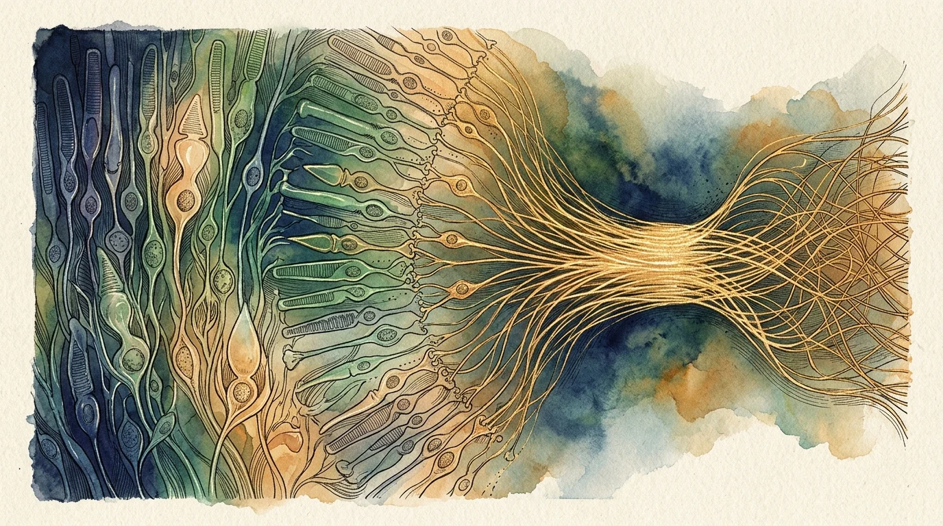
Data from the CDC Vision Health Initiative reveals that over 90 million American adults—representing more than 3 in 5 adults aged 40 and older—live with vision problems or chronic eye conditions. For instance, CDC estimates show that approximately 4.22 million Americans were living with glaucoma in 2022. Alarmingly, 50 percent of individuals with glaucoma do not know they have the disease because early stages produce zero pain or noticeable symptoms.
Research from the National Eye Institute emphasizes that 70 to 90 percent of age-related macular degeneration cases are the “dry” form, which progresses gradually over several years. However, the less common “wet” form causes rapid central vision loss if left untreated. Furthermore, systemic conditions like diabetes double your risk for secondary ocular complications; data shows that 32.2 percent of adults aged 45 and older with diagnosed diabetes have cataracts, 8.6 percent have diabetic retinopathy, 7.1 percent have glaucoma, and 4.3 percent have macular degeneration.

Actionable Strategies for Protecting Long-Term Sight
Prioritize Medical Screening Milestones
The American Academy of Ophthalmology recommends that all adults receive a comprehensive baseline eye examination at age 40 to screen for silent ocular conditions. This baseline exam provides your specialist with a clear benchmark to track subtle anatomical changes over time. For adults aged 65 and older, guidelines from the American Optometric Association recommend scheduling a comprehensive dilated exam every 1 to 2 years, or annually if you manage underlying risk factors like hypertension or diabetes.
Feed Your Eyes with Targeted Ocular Nutrition
Your eyes require specialized antioxidants to filter high-energy blue light and reduce oxidative stress within retinal tissues. Incorporating nutrient-rich dark leafy greens like kale and spinach provides essential carotenoids—lutein and zeaxanthin—that accumulate directly within your macula. Clinical research supported by the National Institutes of Health demonstrates that targeted dietary antioxidants and omega-3 fatty acids from wild-caught fish or flaxseeds strengthen retinal cell membranes and protect small vessel health.
Adopt Daily Lifestyle Habits for Retinal Protection
Protecting your vision extends beyond clinic visits into your daily routine. Wear sunglasses rated for 100 percent UVA and UVB protection whenever you go outdoors, shielding your crystalline lenses from ultraviolet damage that accelerates cataract formation. Managing blood pressure and maintaining steady blood sugar levels protects fragile retinal capillaries from structural damage, while quitting smoking significantly reduces your overall risk for developing advanced macular degeneration.
Real Voices: Clinical Guidance from Eye Health Experts
Optometrists and ophthalmologists frequently remind patients that waiting for visible symptoms to appear before booking an exam poses unnecessary risk. Clinical specialists emphasize that because conditions like early glaucoma and diabetic retinopathy progress without pain or obvious warning signs, proactive screening remains the only reliable method to catch changes before visual fields shrink. Partnering with a trusted eye care professional ensures that subtle shifts in fluid pressure, macular thickness, or lens clarity are identified early, preserving your functional vision and independence for decades.
Frequently Asked Questions About Vision and Aging
How can I tell if my vision changes are normal presbyopia or something more serious?
Normal presbyopia primarily causes difficulty focusing on close print without changing your overall visual clarity or side vision. If you experience distorted lines, dark spots, sudden floaters, or narrowed peripheral vision, you are likely encountering signs of an underlying structural condition rather than normal aging. Schedule a professional exam for an accurate diagnosis.
How often should seniors schedule comprehensive eye examinations?
Medical consensus guidelines recommend that adults aged 65 and older undergo a comprehensive dilated eye exam every 1 to 2 years. If you live with diabetes, high blood pressure, or a family history of eye disease, your doctor may recommend annual screenings to monitor retinal health closely.
Can dietary changes or supplements stop cataracts and macular degeneration?
While nutrition cannot completely cure structural eye disease, consuming foods rich in lutein, zeaxanthin, vitamin C, vitamin E, and zinc supports retinal tissue resilience and slows intermediate macular degeneration. Discuss dietary adjustments and targeted supplement regimens with your eye doctor before starting any new routine.
When should vision changes be treated as an immediate medical emergency?
Seek emergency medical care if you experience sudden vision loss in one or both eyes, a rapid surge in dark floaters with light flashes, a shadow blocking your field of view, or severe eye pain paired with nausea and redness. Immediate clinical intervention can prevent permanent structural blindness.
Taking Your First Step Toward Lasting Sight
Safeguarding your long-term eye health starts with a single, highly manageable action: open your calendar today and schedule a comprehensive baseline or routine eye exam. Reflect on your current visual comfort and write down any subtle changes you have noticed—such as altered night driving vision or frequent prescription updates—to share during your consultation. Taking action now empowers you to protect your sight, maintain your personal autonomy, and enjoy a vibrant quality of life for all the years ahead.








Leave a Reply