

Identifying whether your back pain requires advanced imaging helps you avoid unnecessary radiation while catching critical spinal conditions early. Most episodes resolve with conservative care, yet specific warning signs demand rapid diagnostic scans.
Clinical guidelines show that routine scans for uncomplicated back discomfort often reveal harmless age-related changes that cause needless anxiety. Learning these indicators equips you to seek urgent intervention only when your nerves or structural stability face real threats.
This guide outlines the key red flags that warrant medical imaging alongside evidence-based steps to protect your spine.
Science Snapshot: Understanding Spinal Imaging and Overutilization
Most acute episodes of lumbar discomfort arise from muscular strain, ligamentous irritation, or mild disc inflammation. These tissues typically heal when you combine gentle activity with patience.
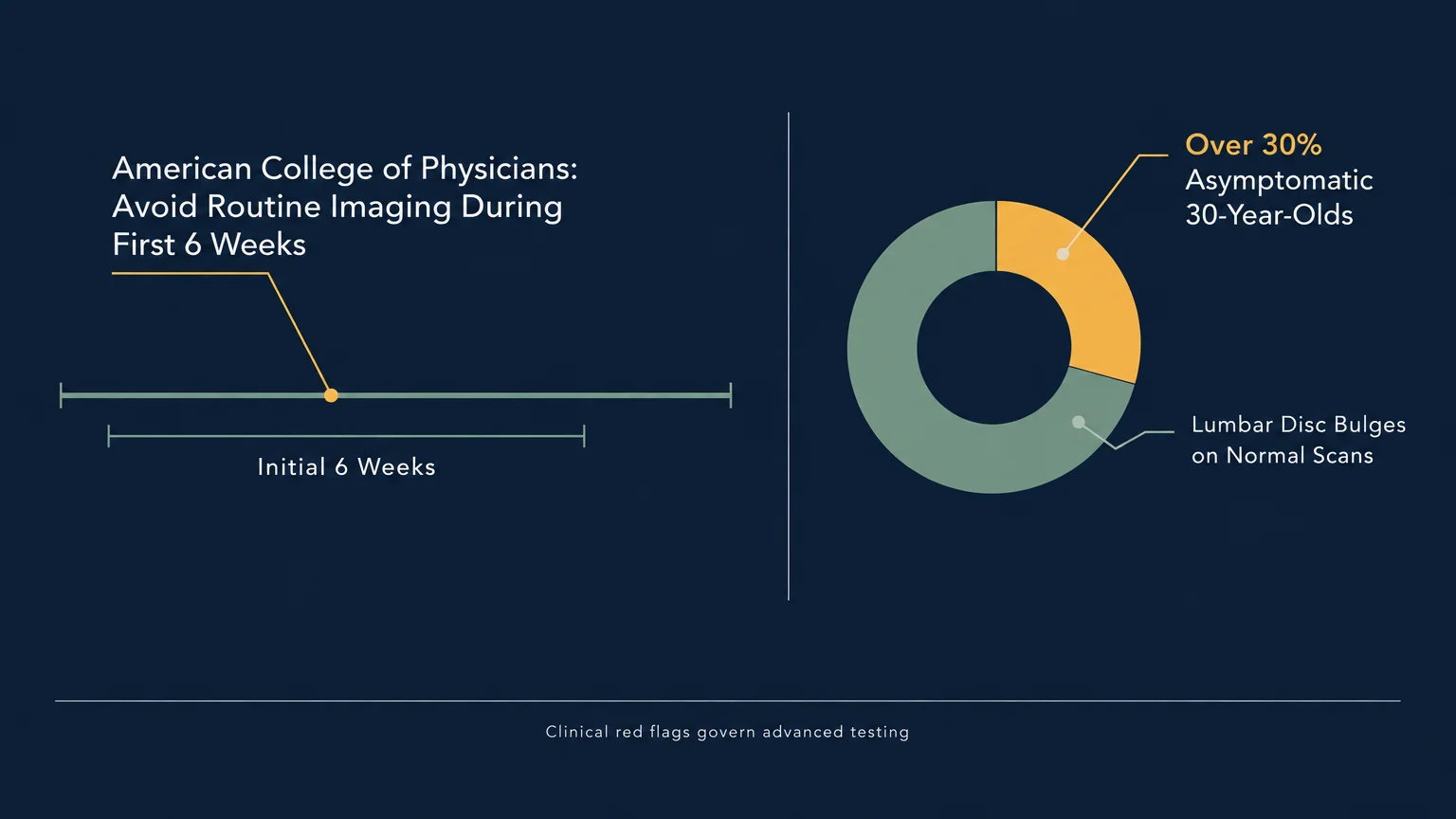
Clinical guidelines from the American College of Physicians recommend avoiding routine imaging during the first six weeks of non-specific lower back pain. Early imaging does not accelerate recovery or improve clinical outcomes for uncomplicated cases.
In fact, healthy adults without back pain regularly exhibit disc bulges, facet joint arthritis, and degenerative changes on normal scans.
Research compiled by the National Institutes of Health reveals that over thirty percent of asymptomatic thirty-year-olds show lumbar disc bulges on magnetic resonance tests.
Unnecessary early scans often lead to invasive procedures, protracted disability, and heightened psychological fear without addressing functional deficits.
Computed tomography and standard X-rays deliver ionizing radiation that requires judicious use, especially in younger individuals.
According to the American Academy of Family Physicians, imaging becomes crucial only when clinical red flags suggest serious structural or systemic pathology.
Knowing when your symptoms warrant advanced testing ensures you receive timely care while bypassing redundant exposure to medical radiation.
8 Symptoms of Back Pain That May Need Imaging
1. Loss of Bowel or Bladder Control
Sudden changes in your bladder or bowel control represent a time-sensitive medical emergency. You should treat any new incontinence or an inability to pass urine as an immediate danger sign.
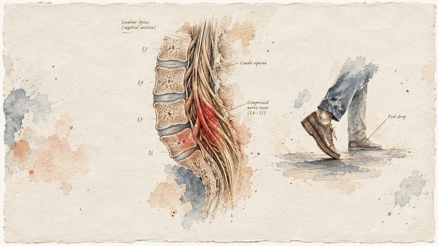
This presentation frequently indicates cauda equina syndrome, where a massive disc herniation or spinal mass compresses the sacral nerve roots. These nerves govern your pelvic organs and lower extremities.
When compressed nerve roots lose blood supply, irreversible neurological impairment can set in within twenty-four to forty-eight hours.
Emergency clinicians utilize emergent magnetic resonance imaging to assess the spinal canal and prepare for rapid surgical decompression.
If you notice urine dribbling, unexpected loss of bowel awareness, or difficulty initiating urination, go to an emergency room immediately.
2. Progressive Neurological Weakness or Foot Drop
If your leg suddenly gives way while walking or your foot drags across the carpet, your motor nerves are facing serious compression.
Clinicians define the inability to lift the front portion of your foot as foot drop. This symptom often stems from severe impingement of the fifth lumbar nerve root.
While mild numbness often improves with time, progressive muscular weakness indicates worsening neurological distress that requires diagnostic precision.
A prompt back pain MRI reveals whether spinal stenosis, structural slipping, or an extruded disc fragment is crushing the motor nerve pathway.
Early surgical decompression for progressive motor loss significantly increases your chances of regaining normal strength and walking mechanics.
3. Spinal Discomfort with a History of Cancer
New or escalating spinal pain in an individual with a personal history of oncology treatment requires prompt diagnostic imaging.
Malignancies affecting the breast, prostate, lung, kidney, or thyroid can metastasize through venous channels directly into your vertebral bones.
These secondary lesions weaken the bony matrix, producing progressive pain and elevating your risk of pathological spinal fractures.
Physicians use contrast-enhanced imaging to distinguish between benign mechanical back strains and malignant structural involvement.
Your care team may coordinate between your oncologist and spine specialist to ensure comprehensive imaging and stable structural support.
4. Unexplained Fever, Chills, or Recent Infection
Spinal pain accompanied by an elevated body temperature, systemic chills, or unprovoked sweats points toward an infectious process.
Bacteria can enter the bloodstream through skin abrasions, dental work, or urinary tract infections and colonize the intervertebral spaces.
This colonization can produce discitis, vertebral osteomyelitis, or an expanding epidural abscess that threatens the adjacent spinal cord.
Immediate diagnostic back pain imaging identifies inflammatory soft-tissue pockets before the infection triggers systemic sepsis or permanent paralysis.
Diagnostic blood tests, including erythrocyte sedimentation rate and C-reactive protein, frequently accompany imaging to assess systemic inflammatory activity.
5. Significant Physical Trauma or Impact
High-impact falls, motor vehicle accidents, or direct blows to your spine require immediate structural evaluation.
For individuals with osteoporosis or those on long-term corticosteroid medication, even minor mechanical jolts can cause vertebral compression fractures.
A timely back pain X-ray provides rapid visualization of spinal curvature, vertebral height loss, and acute bony displacement.
If your doctor suspects subtle bone fragments or spinal cord contusion, computed tomography offers higher resolution across complex bony planes.
Prompt radiological confirmation guides whether conservative bracing, rest, or surgical fixation will best stabilize your injured spine.
6. Saddle Anesthesia Across the Pelvis
A sudden loss of sensation around your groin, buttocks, and inner thighs serves as a major clinical warning sign.
Clinicians call this symptom saddle anesthesia because the numbness aligns with the anatomical areas that contact a riding saddle.
You might notice reduced sensation when sitting, wiping after using the restroom, or engaging in intimate touch.
Saddle anesthesia represents a core component of serious back pain symptoms that demand immediate magnetic resonance imaging to prevent lifelong sensory deficits.
Never wait for sensation to return on its own, as prolonged nerve compression can produce permanent numbness across your pelvic floor.
7. Constant Pain That Wakes You from Sound Sleep
Mechanical back pain usually diminishes when you lie down and find a supported resting posture.
In contrast, unremitting nocturnal pain that awakens you from deep sleep—regardless of how you position pillows—signals possible non-mechanical pathology.
Systemic inflammatory conditions, such as ankylosing spondylitis, as well as occult tumors, often produce deep throbbing that intensifies during overnight rest.
When nocturnal pain persists alongside daytime stiffness, comprehensive imaging helps clinicians identify inflammatory or neoplastic spinal disorders early.
Documenting the exact hours when nighttime pain peaks provides your physician with valuable clues regarding potential inflammatory patterns.
8. Unresponsive Pain After Six Weeks of Conservative Care
Most acute episodes of muscular back pain show measurable improvement within four to six weeks of guided conservative management.
If your discomfort remains disabling despite diligent physical therapy, ergonomic adjustments, and anti-inflammatory support, you need diagnostic clarification.
Evaluating your condition at this stage helps rule out persistent disc tears, unrecognized facet joint arthropathy, or spinal instability.
The resulting imaging findings empower your medical team to customize advanced rehabilitation strategies or evaluate targeted interventional therapies.
A clear scan after six weeks can also reassure you that your spine remains structurally intact, reducing movement-related fear.

Holistic Pillars for Sustainable Spinal Health
Movement: Functional Mechanics and Targeted Decompression
Active movement pumps vital nutrients into your intervertebral discs through passive diffusion, supporting cellular regeneration.
Incorporate short, twenty-minute walks on flat surfaces to lubricate spinal joints and reduce reflexive muscle guarding.
You can also practice low-load core activation exercises, such as the bird-dog and gentle pelvic tilts, to stabilize your lumbar spine.
Adjust your workplace ergonomics by alternating between sitting and standing postures to reduce static compressive pressure on lumbar discs.
Consult a licensed physical therapist to ensure your movement selections match your specific tissue tolerance and rehabilitation stage.
Nutrition: Reducing Systemic and Joint Inflammation
Persistent low-grade inflammation sensitizes peripheral nerves, turning mild structural changes into intense pain experiences.
Build your meals around anti-inflammatory staples such as wild-caught fish, extra virgin olive oil, walnuts, and dark leafy greens.
The World Health Organization highlights balanced nutrition as a cornerstone for preventing long-term musculoskeletal disability and systemic disease.
Limiting ultra-processed foods and refined sugars helps prevent advanced glycation end-products from degrading collagen within intervertebral discs.
Drinking adequate water maintains the hydration of your disc matrices, preserving their shock-absorbing capacity during everyday activities.
Mindset: Calming Central Nervous System Sensitivity
Fear of movement and persistent stress keep your sympathetic nervous system trapped in a state of high alarm.
This heightened arousal causes surrounding back muscles to tighten involuntarily, perpetuating a vicious cycle of pain and tension.
Practicing ten minutes of slow diaphragmatic breathing daily stimulates your vagus nerve and encourages muscular relaxation.
Somatic tracking exercises help you observe bodily sensations with curiosity rather than fear, resetting your brain’s protective danger alarms.
Reframing how you view spinal pain reduces fear, helping your brain interpret bodily sensations with greater calm and accuracy.
Medical Navigation: Partnering with Healthcare Professionals
Navigating back care requires collaborative communication with primary care doctors, chiropractors, and physical therapists.
The Centers for Disease Control and Prevention advocates non-pharmacological therapies as primary care options before considering invasive interventions.
When you discuss back pain red flags with your provider, describe your symptom onset, sensory changes, and functional limits clearly.
Always ask how an imaging scan will alter your treatment roadmap before agreeing to high-cost diagnostic testing.
Voices from the Field: Clinical Perspectives
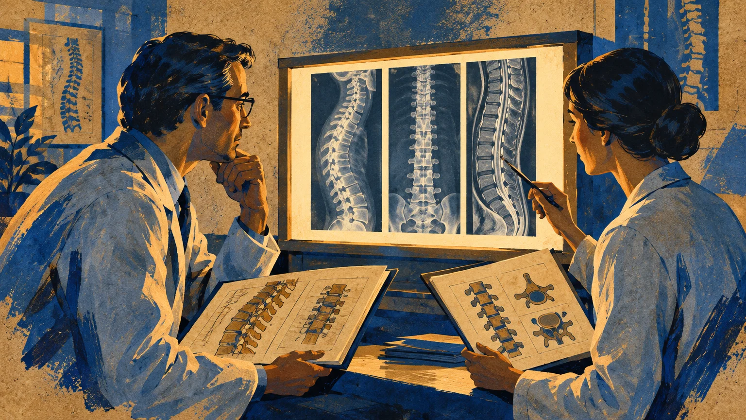
Experienced spinal specialists often emphasize that diagnostic pictures describe your anatomy rather than your functional potential.
Physical therapists frequently observe patients with notable disc bulges who hike, lift weights, and live entirely pain-free lives.
Static diagnostic scans cannot measure muscle endurance, joint mobility, or your nervous system’s capacity to adapt.
Clinicians remind us that active patient participation in physical rehabilitation consistently outperforms passive reliance on imaging scans or injections alone.
Successful recovery relies on treating the whole person through education, restorative movement, and targeted lifestyle modifications.
Frequently Asked Questions
When does back pain need an MRI instead of an X-ray?
An X-ray visualizes bony structures, joint alignment, and fractures with minimal radiation exposure.
An MRI uses magnetic fields to evaluate soft tissues, making it necessary when clinicians suspect disc herniations, nerve compression, or spinal infections.
Your physician will select the appropriate imaging modality based on your specific physical exam findings and clinical history.
Can an MRI definitively explain my back discomfort?
An MRI reveals structural variations, but those variations do not always correlate with where you feel pain.
Many pain-free adults possess asymptomatic disc bulges; therefore, your clinician must correlate scan results with a hands-on physical exam.
What are the most urgent serious back pain symptoms?
Urgent symptoms include loss of bowel or bladder control, saddle anesthesia, progressive foot drop, and spinal pain combined with fever.
If you develop these red flags, proceed to an emergency department or contact a specialist immediately for evaluation.
How long should I wait before seeking diagnostic imaging?
Unless serious red flags appear, guidelines recommend waiting four to six weeks while engaging in active conservative rehabilitation.
If your functional limitations persist or worsen after six weeks, consult your physician to determine whether imaging is appropriate.
Moving Forward with Confidence
Take one practical step today by enjoying a gentle ten-minute walk on flat terrain to nourish your spinal joints.
Observe your physical symptoms with calm awareness, noting whether your mobility is expanding or revealing any red flags.
Your spine is an intrinsically strong structure designed to heal; seek medical guidance whenever unexpected neurological changes arise.








Leave a Reply