

Spotting a new or changing raised mole requires immediate attention, as early detection is the single most effective way to treat potential skin cancer. While most elevated spots are harmless benign growths, understanding the distinct melanoma warning signs allows you to take swift steps to protect your health. Navigating the sea of dermatological information often feels overwhelming, but distinguishing a standard mole from a dangerous lesion is a highly manageable skill. By combining diligent self-examination routines with professional medical guidance, you maintain long-term skin vitality and significantly reduce your risk of serious complications. Knowing exactly what to look for empowers you to prioritize your wellbeing and schedule a timely clinical evaluation without unnecessary panic.
The Science Snapshot: Decoding Your Skin’s Signals
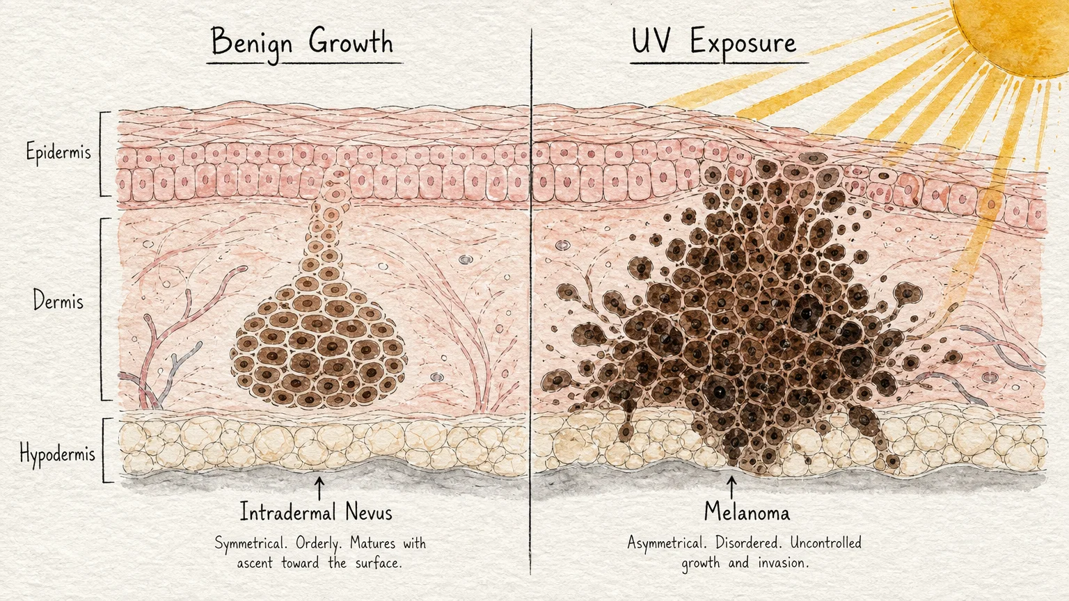
Your skin is a dynamic, living organ that constantly communicates valuable information about your internal health and environmental exposures. Moles, which medical professionals refer to as nevi, develop when pigment-producing cells called melanocytes grow in dense clusters rather than spreading evenly across the surface of your skin. Most individuals develop between ten and forty benign moles throughout their childhood and early adulthood. Over decades, it is entirely biologically appropriate for some of these initially flat spots to elevate slightly, lose their dark pigmentation, and become soft, raised bumps known as intradermal nevi. These harmless changes happen gradually and represent the natural lifecycle of a standard mole maturing as you age.
However, cellular mutations triggered by ultraviolet radiation from the sun or indoor tanning beds can violently disrupt this natural lifecycle, prompting melanocytes to multiply rapidly and erratically. When this rapid cellular division occurs, it can manifest as an unusual raised mole or a firm, dome-shaped nodule that emerges abruptly on previously clear skin. Melanoma, the most aggressive and potentially lethal form of skin cancer, frequently originates in these malfunctioning melanocytes. Other extremely common forms of skin cancer, including basal cell carcinoma and squamous cell carcinoma, also frequently present as elevated, pearly, or crusty bumps that persistently refuse to heal. Reviewing clinical data regarding skin cancer prevalence demonstrates clearly that recognizing these pathological changes in their absolute earliest stages dramatically improves long-term survival rates and minimizes the need for invasive surgical interventions.
Understanding the underlying science behind these cutaneous growths eliminates the crippling mystery and fear often associated with bodily changes. A raised mole is simply a collection of cells taking up vertical space on your epidermis; the crucial medical distinction lies entirely in whether those clustered cells are behaving predictably or proliferating dangerously. By actively learning to read the precise biological signals your skin sends, you shift your entire perspective from passive anxiety to active, informed health management.
Strategy Pillar One: Strategic Observation and the ABCDE Method
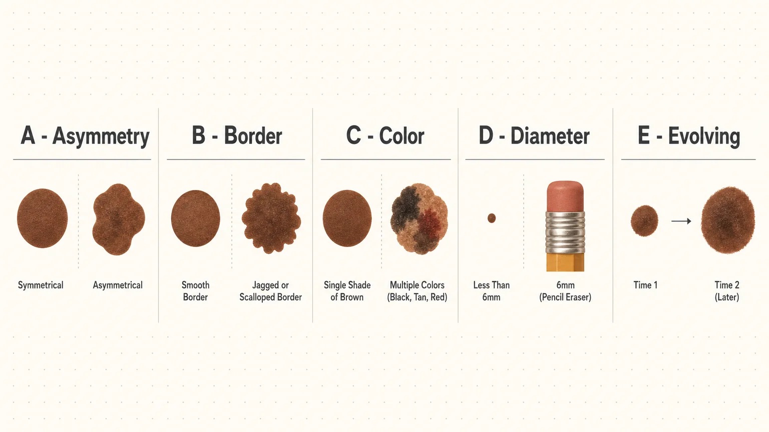
Routine self-examination forms the essential cornerstone of effective, long-term skin health management. Because you observe your own body every single day, you are uniquely positioned to notice the subtle, incremental shifts that a busy doctor might potentially miss during an annual, generalized exam. Dermatologists universally rely on the ABCDE diagnostic framework to evaluate suspicious lesions, and mastering this practical tool enables you to assess a raised mole with remarkable clinical precision right in the comfort of your own home. Rather than rushing through a frantic mirror check, take the deliberate time to evaluate any new or changing elevated spots against these five critical criteria.
Asymmetry and Border Irregularities
A healthy, completely benign mole typically grows symmetrically; if you were to draw an imaginary line directly down its dead center, the two resulting halves would match almost perfectly in volume and shape. When closely examining a raised mole, look specifically for stark asymmetry where one side looks entirely different from the other in overall shape, height, or surface texture. Furthermore, benign moles generally feature smooth, sharp, well-defined borders that clearly and neatly separate the pigmented cells from the surrounding regular skin tissue. Melanoma warning signs frequently include borders that appear noticeably scalloped, jagged, notched, or blurred, occurring as the malignant cells aggressively invade adjacent healthy tissue in a chaotic, haphazard manner.
Color Variations and Diameter Changes
While standard benign moles can range in color from light tan to deep espresso brown, an individual healthy mole typically maintains a uniform, solid, single shade throughout its entire structural composition. A raised mole displaying multiple, distinct colors within the same lesion—such as varying, mottled shades of brown, black, red, white, or even an ominous blue tint—requires immediate, professional evaluation. Diameter also plays a vital diagnostic role; melanomas are frequently larger than six millimeters across, which corresponds roughly to the size of a standard wooden pencil eraser. However, relying solely on size can prove dangerously deceptive, as early-stage melanomas and highly aggressive nodular melanomas can initially present as extremely small raised bumps before expanding rapidly across the skin surface.
Evolution: The Most Crucial Warning Sign
When systematically evaluating a raised mole, evolution represents the single most important, overriding factor you must continually consider. A mole that undergoes sudden, noticeable, or continuous changes in overall size, shape, color, or elevation demands urgent medical attention. Benign moles remain incredibly stable over long periods, whereas cancerous lesions are clinically defined by their rapid, uncontrolled growth and persistent visual transformation. If a previously flat, unassuming mole rapidly becomes heavily elevated, or if an already raised mole suddenly doubles in height, begins to bleed without any provocation, develops a hard crust, or starts to itch intensely, you must entirely bypass home observation and immediately seek clinical care. The physical evolution of a spot always overrides all other traditional diagnostic criteria.
Strategy Pillar Two: Proactive Medical Care and Clinical Screenings
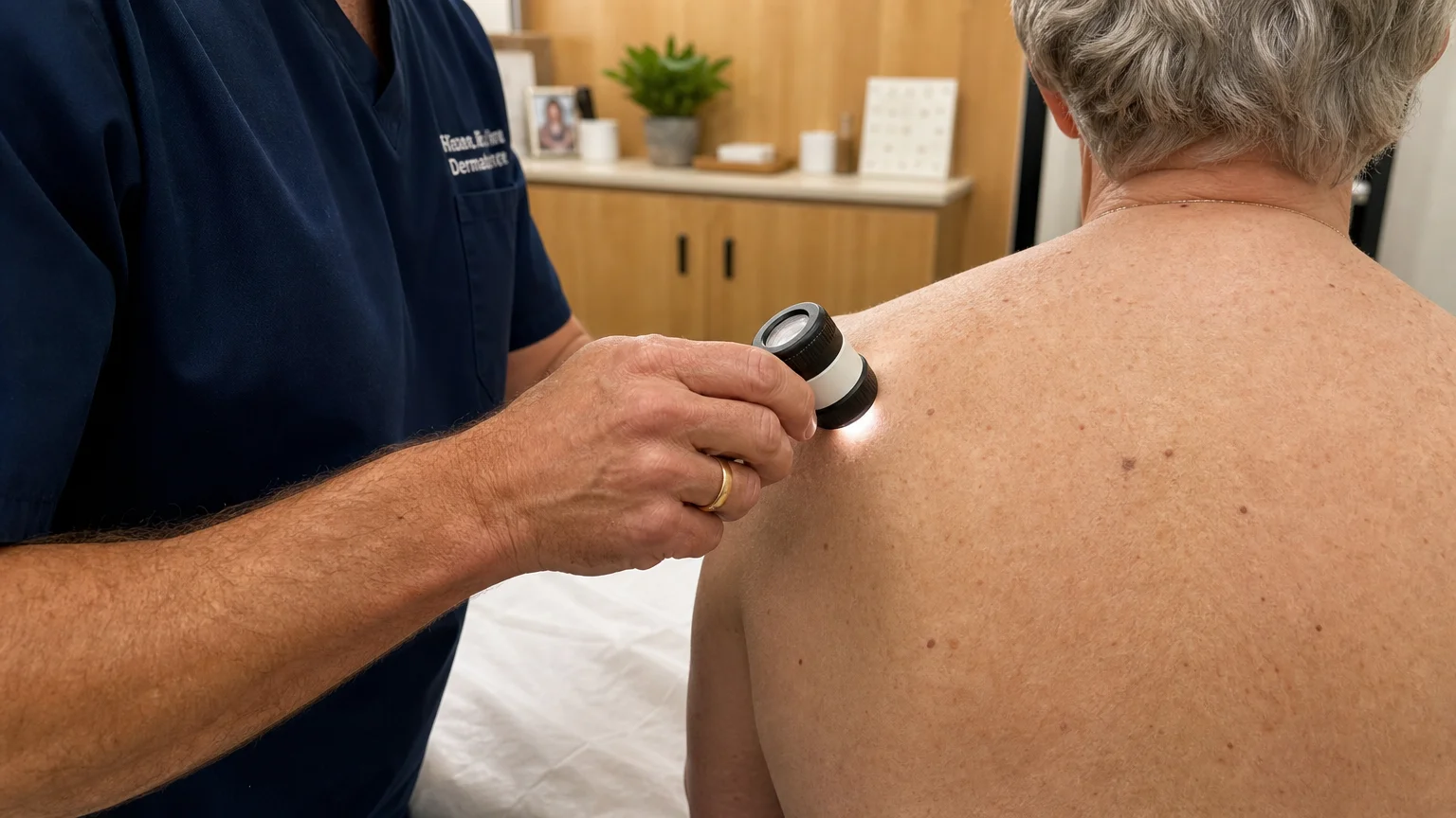
While diligent self-observation empowers your daily routine, establishing an ongoing relationship with a board-certified dermatologist ensures you receive definitive, accurate medical diagnoses. Professional skin cancer screenings involve substantially more than a quick, cursory visual glance from a physician. Modern dermatology specialists utilize an advanced diagnostic tool called a dermatoscope, a specialized instrument that seamlessly combines a powerful magnifying lens with polarized lighting. This sophisticated technology allows the trained clinician to look deeply beneath the very top layer of your skin, revealing intricate pigment networks, microscopic vascular structures, and hidden cellular patterns that remain completely invisible to the naked human eye.
If your dermatologist identifies concerning, irregular features within a raised mole during this examination, they will strongly recommend performing a biopsy. During this incredibly straightforward, outpatient procedure, the physician completely numbs the immediate area with a localized anesthetic and carefully removes the suspicious lesion for comprehensive microscopic laboratory analysis. The standard biopsy process is exceptionally safe, typically takes only a few short minutes to complete, and provides absolute diagnostic certainty for the patient. Receiving a biopsy recommendation naturally provokes anxiety, yet viewing the procedure as an essential, preventative medical tool helps mentally reframe the stressful experience. Securing an accurate pathological diagnosis remains the only definitive way to confirm whether a raised mole cancer sign is genuinely present or if the mysterious growth is entirely benign.
Accessibility to specialized dermatological care unfortunately remains a significant, systemic hurdle for many diverse communities. Factors such as vast geographic isolation, severe lack of specialized local providers, and broader systemic healthcare inequities frequently delay critically important early diagnoses. If securing an appointment with a specialized dermatologist requires enduring an extended, multi-month wait, consulting your primary care physician serves as an excellent, immediate medical alternative. Many dedicated primary care doctors are fully equipped to perform detailed initial evaluations, facilitate necessary punch biopsies, and expedite urgent medical referrals to oncology specialists when they identify pressing melanoma warning signs. You can additionally explore free community skin cancer screening programs that are routinely hosted by regional medical societies and local public health organizations.

Strategy Pillar Three: Holistic Prevention and Long-Term Skin Vitality
Protecting your delicate skin involves committing to comprehensive, daily lifestyle practices that extend far beyond simply checking for suspicious raised spots. Ultraviolet radiation resulting from unprotected sun exposure represents the absolute primary environmental catalyst for the specific cellular mutations that directly lead to skin cancer. Integrating robust, consistent sun protection protocols into your daily morning routine drastically reduces your lifetime risk of developing malignant cutaneous lesions. Applying a premium, broad-spectrum sunscreen with a minimum sun protection factor of thirty every single day—regardless of heavy cloud cover, indoor working conditions, or seasonal temperature changes—provides an essential, non-negotiable defensive barrier for your vulnerable epidermis.
Protective clothing frequently offers an equally critical, and often substantially more reliable, method of physically shielding your skin from harm. Broad-brimmed sun hats, wraparound sunglasses featuring complete ultraviolet protection, and specialized garments manufactured with designated ultraviolet protection factor fabrics physically block dangerous radiation rays from ever penetrating your dermal cells. Furthermore, actively seeking dense shade during the sun’s absolute peak intensity hours between ten in the morning and four in the afternoon drastically cuts your cumulative daily radiation exposure. You must also entirely avoid the dangerous use of artificial tanning beds, as the highly concentrated ultraviolet radiation they actively emit exponentially increases the statistical likelihood of melanoma development, particularly when frequently utilized during vulnerable adolescence and early adulthood.
From a deeply holistic standpoint, aggressively supporting your skin’s innate cellular resilience through a highly nutrient-dense diet also plays a valuable, supportive role in maintaining long-term bodily vitality. Consuming a diverse, colorful array of fresh vegetables, whole fruits, and lean proteins actively provides your body with the potent, essential antioxidants required to effectively combat internal oxidative stress and rapidly repair minor cellular damage caused by unavoidable daily environmental exposures. While no specific, isolated food can miraculously prevent or cure skin cancer, maintaining optimal systemic health heavily ensures your complex immune system functions efficiently, potentially assisting your resilient body in identifying and neutralizing abnormal, mutated cells long before they aggressively develop into a concerning raised mole. Understanding the broad, medically sound preventative guidelines for skin safety helps you successfully build a resilient, highly sustainable lifestyle that naturally safeguards your overall dermal health.

Real Voices: Clinical Insights on Raised Spots
Medical professionals continuously and passionately advocate for highly proactive patient involvement in personal dermatological health. Leading clinical oncologists and practicing dermatologists frequently emphasize the supreme importance of quickly identifying the proverbial ugly duckling on your skin landscape. This practical diagnostic concept suggests that standard benign moles on a single individual typically resemble one another heavily, sharing highly similar baseline colors, basic shapes, and general sizes. A newly discovered raised mole that looks profoundly and obviously different from all the surrounding benign spots—the ugly duckling—warrants immediate, professional scrutiny, even if it does not strictly meet all the traditional, established ABCDE diagnostic criteria.
Clinicians also heavily stress the absolute, critical necessity of inclusive, broad skin cancer awareness. A pervasive and highly dangerous societal misconception persists that deadly skin cancer exclusively afflicts individuals possessing very fair skin and light hair. In stark medical reality, melanoma and other aggressive skin cancers deeply affect people of all racial backgrounds, ethnic origins, and varying skin tones. When severe skin cancer initially develops in individuals possessing deeply pigmented skin, it is frequently diagnosed at a much later, vastly more dangerous stage precisely because both unsuspecting patients and under-trained medical practitioners completely overlook the subtle early warning signs. Dermatologists urgently press everyone, completely regardless of their natural complexion, to diligently monitor their entire bodies for new growths, paying incredibly special attention to easily neglected, hidden areas such as the palms of the hands, the soles of the feet, and directly beneath the fingernails. Reviewing comprehensive, culturally inclusive dermatology resources tailored for people of color highlights the absolute, non-negotiable necessity of universal vigilance and comprehensive, equitable clinical care for all populations.
Frequently Asked Questions
Do all raised moles need to be surgically removed?
Absolutely not. The vast, overwhelming majority of standard raised moles are completely harmless, benign intradermal nevi that pose zero actual threat to your overall systemic health. Certified dermatologists generally strongly advise against surgically removing completely healthy, benign growths unless they cause you significant, ongoing physical discomfort—such as repeatedly catching painfully on your restrictive clothing or heavy jewelry—or if you specifically desire professional removal for entirely personal cosmetic reasons. Completely unnecessary surgical removal inevitably results in permanent localized skin scarring and carries standard, unavoidable procedural risks like minor bacterial infections. Your treating physician will only strictly mandate a surgical excision if the specific raised mole clearly exhibits highly suspicious visual features, undergoes rapid sudden changes, or returns a definitively positive laboratory biopsy result actively indicating malignant cellular activity.
Can a perfectly flat mole evolve into a raised mole over time naturally?
Yes, slowly transitioning from a flat, unassuming spot to a soft, raised bump actually represents a perfectly normal, biologically expected evolutionary pathway for many standard, harmless moles. As you naturally age, the dense, pigmented clusters of melanocytes that initially form a distinct mole can gradually sink much deeper into the underlying dermal layers of your skin, naturally causing the visible surface tissue to elevate slightly, soften considerably, and frequently lose its original dark brown pigmentation. This incredibly natural maturation process occurs extraordinarily slowly, typically spanning multiple decades of your life. The genuine medical concern only arises when a previously flat spot elevates incredibly rapidly over a highly compressed period of a few short weeks or months, which strongly indicates chaotic, dangerous cellular division requiring immediate clinical investigation.
Does a raised mole that occasionally bleeds or itches always indicate skin cancer?
While spontaneous bleeding, unexpected oozing, and persistent, nagging itching absolutely represent highly recognized melanoma warning signs, experiencing these alarming symptoms does not automatically guarantee a devastating cancer diagnosis. Because a standard raised mole physically protrudes above the surrounding flat skin, it frequently sustains highly localized, minor physical trauma from everyday mechanical friction caused by tight pant waistbands, restrictive bra straps, or simple accidental scratching. This entirely routine physical irritation easily causes a completely harmless mole to rapidly become inflamed, intensely itchy, or highly prone to minor, superficial bleeding. However, you should absolutely never blindly assume that simple clothing friction is the sole culprit; any bothersome lesion that repeatedly bleeds, actively develops a persistent hard crust, or totally fails to heal completely within three short weeks requires an immediate, thorough professional medical evaluation to definitively rule out underlying malignancy.
How frequently should I perform a comprehensive skin self-exam at home?
Top dermatologists strongly and uniformly recommend performing a highly thorough, full-body skin self-examination exactly once per month. Systematically checking your skin monthly allows you to become deeply, intimately familiar with the completely unique baseline topography of your existing moles, making it significantly easier to confidently spot remarkably subtle new growths or concerning evolutionary changes. Conducting the examination vastly more frequently than once a month often leads to severely heightened health anxiety and makes microscopic, completely normal day-to-day bodily changes incredibly difficult to accurately track and assess. Always ensure you meticulously perform your personal self-exam in a brilliantly lit room, specifically utilizing both a large full-length mirror and a small handheld mirror to meticulously inspect historically difficult-to-see areas like your mid-back, the very back of your neck, and the complete posterior of your legs.
Take Your Next Confident Step
Protecting your vital skin organ requires consistent, deliberate action rather than harboring overwhelming, paralyzing anxiety. If you have recently noticed a new or actively changing raised mole on your body, your most highly constructive immediate step involves precise photographic documentation. Find a brilliantly lit area today and use your personal smartphone to capture a highly clear, sharply focused, macro photograph of the specific spot in question, carefully placing a standard coin or a small ruler directly next to the lesion to firmly establish an accurate, undeniable scale for its exact current diameter. This critical baseline image immediately provides your consulting dermatologist with invaluable, concrete historical data to accurately evaluate any future evolutionary changes. Always remember that highly proactive home monitoring coupled with incredibly swift professional medical intervention remain your absolute most powerful tools for maintaining lasting, vibrant skin health and ensuring total peace of mind.


Leave a Reply